File:Integumentary- hair follicle 01.jpg: Difference between revisions
No edit summary |
|||
| Line 10: | Line 10: | ||
See also [[:File:Integumentary- hair follicle 02.jpg|Hair follicle alternate labelling]] | See also [[:File:Integumentary- hair follicle 02.jpg|Hair follicle alternate labelling]] | ||
{{Hair histology links}} | |||
(Text modified from UWA Blue Histology) | (Text modified from UWA Blue Histology) | ||
Revision as of 14:30, 23 February 2013
Integumentary - Hair Follicle Histology
(Stain - Haematoxylin Eosin)
- Hair follicles of terminal hair span the entire dermis and usually extend deep into the hypodermis.
- Most of them will be cut at odd angles and only a few good longitudinally or transversely cut profiles are visible.
- The hair may have been lost during the preparation of the specimen and not all hair follicles will contain hairs.
- Although it is often possible to see the attachment of the arrector pili muscle into the hair follicle or the papillary layer of the dermis, both attachments are hardly ever visible in the same section.
See also Hair follicle alternate labelling
- Hair Links: Follicle in skin 1 | Follicle in skin 2 | Follicle label 1 | Follicle label 2 | Sebaceous gland 1 | Sebaceous gland 2
Links: Histology | Histology Stains | Blue Histology images copyright Lutz Slomianka 1998-2009. The literary and artistic works on the original Blue Histology website may be reproduced, adapted, published and distributed for non-commercial purposes. See also the page Histology Stains.
Cite this page: Hill, M.A. (2024, June 2) Embryology Integumentary- hair follicle 01.jpg. Retrieved from https://embryology.med.unsw.edu.au/embryology/index.php/File:Integumentary-_hair_follicle_01.jpg
- © Dr Mark Hill 2024, UNSW Embryology ISBN: 978 0 7334 2609 4 - UNSW CRICOS Provider Code No. 00098G
(Text modified from UWA Blue Histology)
Links: Histology | Histology Stains | Blue Histology images copyright Lutz Slomianka 1998-2009. The literary and artistic works on the original Blue Histology website may be reproduced, adapted, published and distributed for non-commercial purposes. See also the page Histology Stains.
Cite this page: Hill, M.A. (2024, June 2) Embryology Integumentary- hair follicle 01.jpg. Retrieved from https://embryology.med.unsw.edu.au/embryology/index.php/File:Integumentary-_hair_follicle_01.jpg
- © Dr Mark Hill 2024, UNSW Embryology ISBN: 978 0 7334 2609 4 - UNSW CRICOS Provider Code No. 00098G
File history
Click on a date/time to view the file as it appeared at that time.
| Date/Time | Thumbnail | Dimensions | User | Comment | |
|---|---|---|---|---|---|
| current | 13:34, 26 March 2012 | 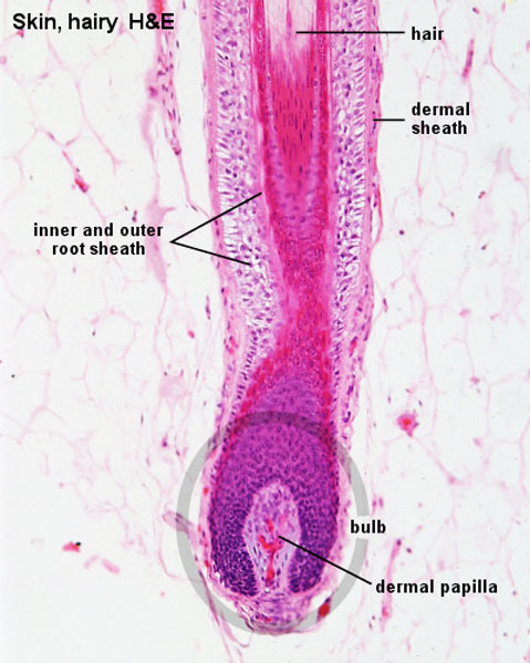 | 479 × 599 (66 KB) | Z8600021 (talk | contribs) | |
| 14:27, 13 October 2010 | 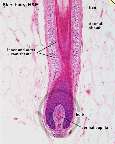 | 400 × 500 (75 KB) | S8600021 (talk | contribs) | ==Integumentary- hair follicle histology== Hair follicles of terminal hair span the entire dermis and usually extend deep into the hypodermis. Most of them will be cut at odd angles and only a few good longitudinally or transversely cut profiles are visi |
You cannot overwrite this file.
File usage
The following 4 pages use this file: