File:Wilms tumor.jpg
Wilms_tumor.jpg (776 × 512 pixels, file size: 310 KB, MIME type: image/jpeg)
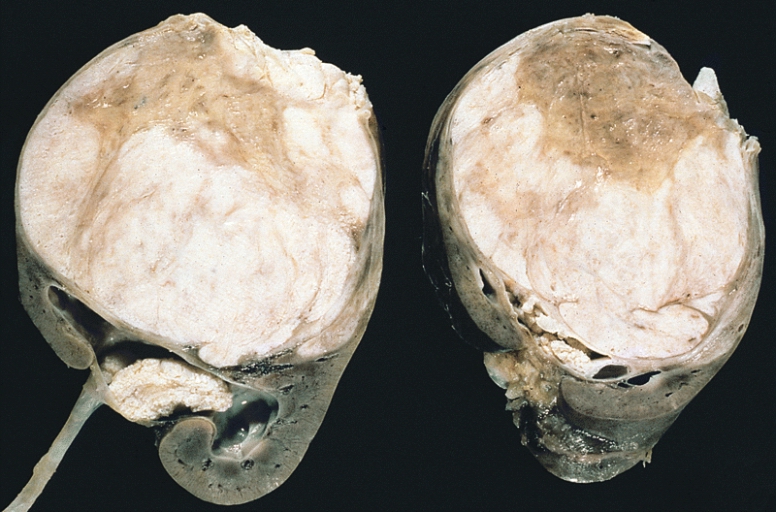
Renal Nephroblastoma
KIDNEY-BLADDER-URINARY: NEPHROBLASTOMA
Note the prominent septa subdividing the sectioned surface and the protrusion of tumor into the renal pelvis, resembling botryoid rhabdomyosarcoma.
Reference
Date 20 March 2008
Image and description are from the AFIP Atlas of Tumor Pathology, according to entry #407018 in Pathology Education Instructional Resource. The Armed Forces Institute of Pathology Electronic Fascicles (CD-ROM Version of the Atlas of Tumor Pathology) contains U.S. Government work which may be used without restriction.
Author: The Armed Forces Institute of Pathology
This work is in the public domain in the United States because it is a work of the United States Federal Government under the terms of Title 17, Chapter 1, Section 105 of the US Code.
http://commons.wikimedia.org/wiki/File:Wilms_tumor.jpg
Cite this page: Hill, M.A. (2024, April 23) Embryology Wilms tumor.jpg. Retrieved from https://embryology.med.unsw.edu.au/embryology/index.php/File:Wilms_tumor.jpg
- © Dr Mark Hill 2024, UNSW Embryology ISBN: 978 0 7334 2609 4 - UNSW CRICOS Provider Code No. 00098G
File history
Click on a date/time to view the file as it appeared at that time.
| Date/Time | Thumbnail | Dimensions | User | Comment | |
|---|---|---|---|---|---|
| current | 14:02, 19 September 2009 |  | 776 × 512 (310 KB) | S8600021 (talk | contribs) | KIDNEY-BLADDER-URINARY: NEPHROBLASTOMA Note the prominent septa subdividing the sectioned surface and the protrusion of tumor into the renal pelvis, resembling botryoid rhabdomyosarcoma. Date 20 March 2008 Source Image and description are from the AF |
You cannot overwrite this file.
File usage
The following 4 pages use this file:
