File:West02.jpg
West02.jpg (619 × 549 pixels, file size: 34 KB, MIME type: image/jpeg)
Fig. 2
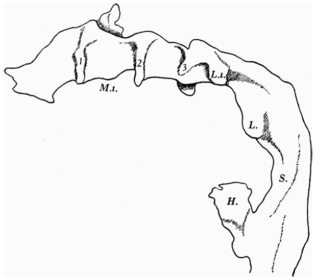
The pharynx, as a whole (Text-fig. 2), is concave dorsally from side to side on its dorsal surface and concave ventrally on its ventral surface.
The floor is marked in its most anterior part by a deep median groove, and a much shallower groove likewise marks the dorsal wall, or roof. At the point of attachment of the pharyngeal membrane the groove on the roof of the pharynx is continued on to the roof of the mouth, and there it divides into two limbs with a rounded ridge between them; this ridge is due to the down growth of the forebrain and emphasises the close relationship between brain and pharynx at this point; just to the cranial side of the pharyngeal membrane the pouch of Rathke shows as a small diverticulum of the mouth cavity; Seessel's pouch cannot be identified.
| Historic Disclaimer - information about historic embryology pages |
|---|
| Pages where the terms "Historic" (textbooks, papers, people, recommendations) appear on this site, and sections within pages where this disclaimer appears, indicate that the content and scientific understanding are specific to the time of publication. This means that while some scientific descriptions are still accurate, the terminology and interpretation of the developmental mechanisms reflect the understanding at the time of original publication and those of the preceding periods, these terms, interpretations and recommendations may not reflect our current scientific understanding. (More? Embryology History | Historic Embryology Papers) |
- Links: Fig 1 | Fig 2 | Fig 3 | Fig 4 | Fig 5 | Fig 6 | Fig 7 | Fig 8 | Fig 9 | Fig 10 | Fig 11 | Plate 1 | Plate 1 Fig 1 | Plate 1 Fig 2 | Plate 1 Fig 3 | Plate 1 Fig 4 | Plate 1 Fig 5 | Plate 1 Fig 6
Reference
West CM. A human embryo of twenty-five somites. (1937) J. Anat., 71(2): 169-200.1. PMID 17104635
Cite this page: Hill, M.A. (2024, April 25) Embryology West02.jpg. Retrieved from https://embryology.med.unsw.edu.au/embryology/index.php/File:West02.jpg
- © Dr Mark Hill 2024, UNSW Embryology ISBN: 978 0 7334 2609 4 - UNSW CRICOS Provider Code No. 00098G
File history
Click on a date/time to view the file as it appeared at that time.
| Date/Time | Thumbnail | Dimensions | User | Comment | |
|---|---|---|---|---|---|
| current | 15:20, 28 January 2012 |  | 619 × 549 (34 KB) | S8600021 (talk | contribs) | ==Fig. 2 == {{Template:West1937}} {{Historic Disclaimer}} {{Historic Papers}} ===Reference=== <pubmed>17104635</pubmed>| [http://www.ncbi.nlm.nih.gov/pmc/articles/PMC1252340 PMC1252340] Category:Carnegie Stage 12 |
You cannot overwrite this file.
File usage
The following page uses this file:

