File:Vein histology 01.jpg
Vein_histology_01.jpg (480 × 600 pixels, file size: 57 KB, MIME type: image/jpeg)
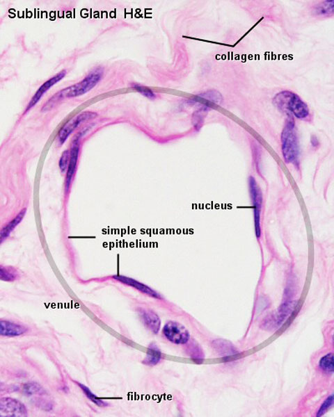
Vein Histology
The vein endothelium is an example of a simple squamous epithelium. (Stain - Haematoxylin Eosin)
- single layer of flattened, scale-like or plate-like cells.
- large body cavities and heart, blood vessels and lymph vessels are typically lined by a simple squamous epithelium.
- nuclei of the epithelial cells are often flattened or ovoid, and they are located close to the centre of the cells.
Note the entire cardiovascular system is lined by this type of epithelium.
- Links: Blood Vessel Development
Links: Histology | Histology Stains | Blue Histology images copyright Lutz Slomianka 1998-2009. The literary and artistic works on the original Blue Histology website may be reproduced, adapted, published and distributed for non-commercial purposes. See also the page Histology Stains.
Cite this page: Hill, M.A. (2024, April 23) Embryology Vein histology 01.jpg. Retrieved from https://embryology.med.unsw.edu.au/embryology/index.php/File:Vein_histology_01.jpg
- © Dr Mark Hill 2024, UNSW Embryology ISBN: 978 0 7334 2609 4 - UNSW CRICOS Provider Code No. 00098G
File history
Click on a date/time to view the file as it appeared at that time.
| Date/Time | Thumbnail | Dimensions | User | Comment | |
|---|---|---|---|---|---|
| current | 13:03, 26 March 2012 | | 480 × 600 (57 KB) | Z8600021 (talk | contribs) | ==Vein Histology== The vein endothelium is an example of a simple squamous epithelium. {{Blue Histology}} |
You cannot overwrite this file.