File:Ultrasound uterine and ovarian vascularity.jpg

Original file (531 × 780 pixels, file size: 132 KB, MIME type: image/jpeg)
Ultrasound uterine and ovarian vascularity
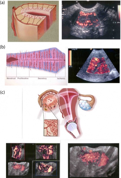
Two-dimensional vs three-dimensional transvaginal color Doppler sonography (TV-CDS).
(a) Diagram (left) and two-dimensional TV-CDS (right) showing arcuate, radial, and spiral vessels in follicular phase.
(b) Diagram (left) are two-dimensional TV-CDS (right) showing changes in endometrial vascularity throughout the menstral cycle. During the luteal phase, several spiral vessels are detected.
(c) Diagram (top) and multiplanar images (left) and magnified 3D TV-CDS (right) of corpus luteum. The multiplanar image shows the vascular "wreath" surrounding the functioning corpus luteum in the longitudinal (top left), short (axial) (top right) and coronal (bottom right) planes. The combined gray scale and 3D TV-CDS image (both left) which is also magnified and shown as the right depicts the numerous vessels surrounding this corpus luteum.
- Links: Ultrasound | Menstrual Cycle | Ovary Development | Uterus Development
Reference
<pubmed>16095530</pubmed>| PMC1208937 | J. Exp. Clin. Assist.
Copyright
© 2005 Raine-Fenning and Fleischer; licensee BioMed Central Ltd.
This is an Open Access article distributed under the terms of the Creative Commons Attribution License (http://creativecommons.org/licenses/by/2.0), which permits unrestricted use, distribution, and reproduction in any medium, provided the original work is properly cited.
Original File Name: Figure 1 1743-1050-2-10-1.jpg http://www.ncbi.nlm.nih.gov/pmc/articles/PMC1208937/figure/F1
Paper Abstract
- "This overview describes and illustrates the clinical applications of three-dimensional transvaginal sonography in reproductive medicine. Its main applications include assessment of uterine anomalies, intrauterine pathology, tubal patency, polycystic ovaries, ovarian follicular monitoring and endometrial receptivity. It is also useful for detailed evaluation of failed and/or ectopic pregnancy. Three-dimensional color Doppler sonography provides enhanced depiction of uterine, endometrial, and ovarian vascularity."
File history
Click on a date/time to view the file as it appeared at that time.
| Date/Time | Thumbnail | Dimensions | User | Comment | |
|---|---|---|---|---|---|
| current | 12:54, 30 July 2011 |  | 531 × 780 (132 KB) | S8600021 (talk | contribs) | ==Ultrasound uterine and ovarian vascularity== Two-dimensional vs Three-dimensional transvaginal color Doppler sonography (a) Diagram (left) and two-dimensional TV-CDS (right) showing arcuate, radial, and spiral vessels in follicular phase. (b) Diagram |
You cannot overwrite this file.
File usage
The following 4 pages use this file: