File:Supernumerary renal vein 03.jpg

Original file (800 × 794 pixels, file size: 80 KB, MIME type: image/jpeg)
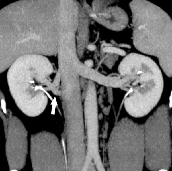
Supernumerary Renal Vein
- most common venous anomalies are multiple renal veins seen in approximately 15-30% of patients
- more common on the right side and these occur in up to 30% of individuals
- Supernumerary right renal vein in 30-year-old male voluntary kidney donor.
- Curved coronal maximum intensity projection image show two right renal veins crossing each other and draining into inferior vena cava (arrows).
Renal Vascular Anomalies: Multiple renal arteries | Accessory renal artery | Supernumerary right renal vein 1 | Supernumerary right renal vein 1 | Multiple right renal veins 2 | Multiple right renal veins 2 | Cardiovascular System Development
Original file name: Fig. 8 kjr-11-346-g008.jpg (panel A cropped from full image)
Reference
Kumar S, Neyaz Z & Gupta A. (2010). The utility of 64 channel multidetector CT angiography for evaluating the renal vascular anatomy and possible variations: a pictorial essay. Korean J Radiol , 11, 346-54. PMID: 20461189 DOI.
Copyright
This is an Open Access article distributed under the terms of the Creative Commons Attribution Non-Commercial License (http://creativecommons.org/licenses/by-nc/3.0) which permits unrestricted non-commercial use, distribution, and reproduction in any medium, provided the original work is properly cited.
Cite this page: Hill, M.A. (2024, April 16) Embryology Supernumerary renal vein 03.jpg. Retrieved from https://embryology.med.unsw.edu.au/embryology/index.php/File:Supernumerary_renal_vein_03.jpg
- © Dr Mark Hill 2024, UNSW Embryology ISBN: 978 0 7334 2609 4 - UNSW CRICOS Provider Code No. 00098G
File history
Click on a date/time to view the file as it appeared at that time.
| Date/Time | Thumbnail | Dimensions | User | Comment | |
|---|---|---|---|---|---|
| current | 12:31, 3 September 2011 | | 800 × 794 (80 KB) | S8600021 (talk | contribs) | ==Supernumerary Renal Vein== * most common venous anomalies are multiple renal veins seen in approximately 15-30% of patients * more common on the right side and these occur in up to 30% of individuals * Supernumerary right renal vein in 30-year-old male |
You cannot overwrite this file.
File usage
The following 3 pages use this file: