File:Stage7 bf5a.jpg
From Embryology
Size of this preview: 800 × 563 pixels. Other resolution: 1,024 × 721 pixels.
Original file (1,024 × 721 pixels, file size: 690 KB, MIME type: image/jpeg)
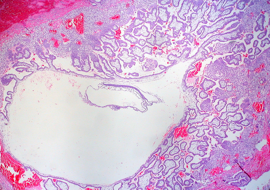
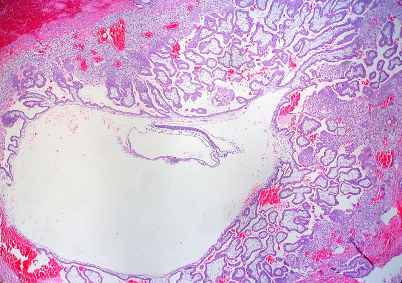
Human Embryo Stage 7
Appearance of section through implanted conceptus appears to be about a Carnegie stage 7 embryo. Photo author states similar to the Hertig-Rock embryo (1945) dated as 16.5 to 19 days post-ovulation, or GA Week 4 to 5 week.
- Features: embryonic disc, primitive node, primative streak, primitive groove, yolk sac
- Facts: Week 3, 15 - 17 days, 0.4 mm
- View: embryonic disc and chorionic vesicle.
- Events: Gastrulation is continuing as cells migrate from the epiblast, continuing to form mesoderm.
- Stage 7 Links: Large image | Medium image | Small image | Trilaminar embryo excerpt | Villi excerpt 1 | Villi excerpt 2 | Carnegie stage 7
- Carnegie Stages: 1 | 2 | 3 | 4 | 5 | 6 | 7 | 8 | 9 | 10 | 11 | 12 | 13 | 14 | 15 | 16 | 17 | 18 | 19 | 20 | 21 | 22 | 23 | About Stages | Timeline
Image: Dr Ed Uthman (Houston, Texas) - other pathology images - CC BY 2.0
Cite this page: Hill, M.A. (2024, April 18) Embryology Stage7 bf5a.jpg. Retrieved from https://embryology.med.unsw.edu.au/embryology/index.php/File:Stage7_bf5a.jpg
- © Dr Mark Hill 2024, UNSW Embryology ISBN: 978 0 7334 2609 4 - UNSW CRICOS Provider Code No. 00098G
File history
Click on a date/time to view the file as it appeared at that time.
| Date/Time | Thumbnail | Dimensions | User | Comment | |
|---|---|---|---|---|---|
| current | 14:44, 21 July 2010 | | 1,024 × 721 (690 KB) | S8600021 (talk | contribs) | ==Human embryo - 3 week== Appearance of section looks to be about a Carnegie stage 7 embryo. Original Author Legend - Primitive Trilaminar Human Embryo in Tubal Pregnancy (40X) :"I think this is at about the same developmental stage as the Hertig-Roc |
You cannot overwrite this file.
File usage
There are no pages that use this file.