File:Spermatozoa tail EM01.jpg

Original file (932 × 613 pixels, file size: 75 KB, MIME type: image/jpeg)
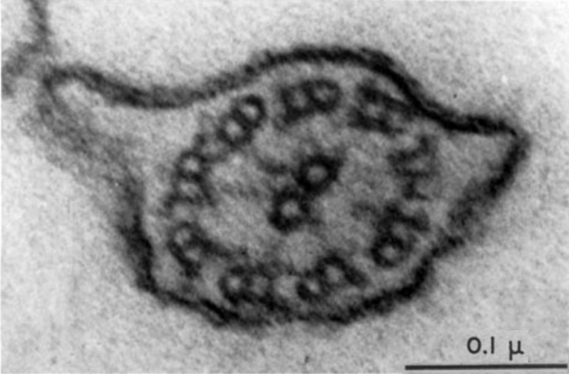
Cross-section through a sperm tail
The features visible here are the same as those shown in Fig. 1 or in Text-fig. 1.
The cell membrane can be seen as a "double membrane" at many places.
X 240,000.
This image is from an historic paper that identified features of the spermatozoa microtubule organisation by EM.
References
<pubmed>13654448</pubmed>| PMC2224653 | PDF
Copyright
Rockefeller University Press - Copyright Policy This article is distributed under the terms of an Attribution–Noncommercial–Share Alike–No Mirror Sites license for the first six months after the publication date (see http://www.jcb.org/misc/terms.shtml). After six months it is available under a Creative Commons License (Attribution–Noncommercial–Share Alike 4.0 Unported license, as described at https://creativecommons.org/licenses/by-nc-sa/4.0/ ). (More? Help:Copyright Tutorial)
Fig.3 PMID 13654448 Original image cropped, contrast adjusted rotated and labelling moved.
File history
Click on a date/time to view the file as it appeared at that time.
| Date/Time | Thumbnail | Dimensions | User | Comment | |
|---|---|---|---|---|---|
| current | 06:21, 16 March 2012 |  | 932 × 613 (75 KB) | Z8600021 (talk | contribs) | === References === <pubmed>13654448</pubmed>| [http://www.ncbi.nlm.nih.gov/pmc/articles/PMC2224653/pdf/269.pdf PDF] {{JCB}} Text Fig.1 PMID 13654448 Category:Spermatozoa Category:Sea Urchin Category:Electron Micrograph |
You cannot overwrite this file.
File usage
The following page uses this file: