File:Skeletal muscle histology 007.jpg
From Embryology
Size of this preview: 750 × 600 pixels. Other resolution: 1,280 × 1,024 pixels.
Original file (1,280 × 1,024 pixels, file size: 253 KB, MIME type: image/jpeg)
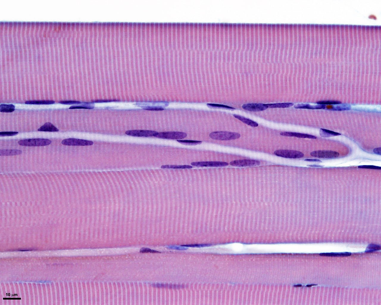
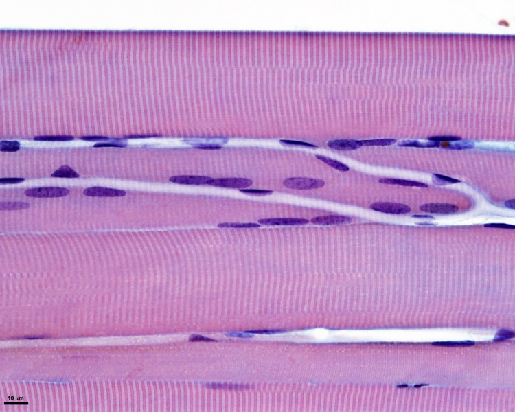
Skeletal Muscle Histology
- Human skeletal muscle
- longitudinal section of muscle fibres
- capillaries visible
- Stain Whipf's polychrome magnification x40
- Muscle Histology: Muscle Development | Human HE x4 longitudinal and transverse | Human HE x40 transverse | Human HE x40 longitudinal | Human HE x40 longitudinal | Human HE x4 longitudinal and transverse | Muscle Spindle HE x40 | Human HE x40 | Human HE x40 | Human HE x40 | Human HE x100 | Human HE x100 | Fetal human muscle | Myotendinous junction label | Myotendinous junction HE x40 | Whipf 1 | Whipf 2 | Whipf 3 | Tongue HE x10 transverse | Tongue x100 | Muscle spindle HE x20 | Muscle spindle HE x40
Links: Histology | Histology Stains | Blue Histology images copyright Lutz Slomianka 1998-2009. The literary and artistic works on the original Blue Histology website may be reproduced, adapted, published and distributed for non-commercial purposes. See also the page Histology Stains.
Cite this page: Hill, M.A. (2024, April 18) Embryology Skeletal muscle histology 007.jpg. Retrieved from https://embryology.med.unsw.edu.au/embryology/index.php/File:Skeletal_muscle_histology_007.jpg
- © Dr Mark Hill 2024, UNSW Embryology ISBN: 978 0 7334 2609 4 - UNSW CRICOS Provider Code No. 00098G
File history
Click on a date/time to view the file as it appeared at that time.
| Date/Time | Thumbnail | Dimensions | User | Comment | |
|---|---|---|---|---|---|
| current | 17:02, 2 October 2011 | | 1,280 × 1,024 (253 KB) | S8600021 (talk | contribs) | ==Human Skeletal Muscle Histology== * Human skeletal muscle * longitudinal section of muscle fibres * capillaries visible * Stain Whipf's polychrome magnification x40 {{SkMhistolinks}} {{Blue Histology}} Category:Human |
You cannot overwrite this file.
File usage
The following 3 pages use this file: