File:Single human spermatozoa.jpg

Original file (1,000 × 780 pixels, file size: 53 KB, MIME type: image/jpeg)
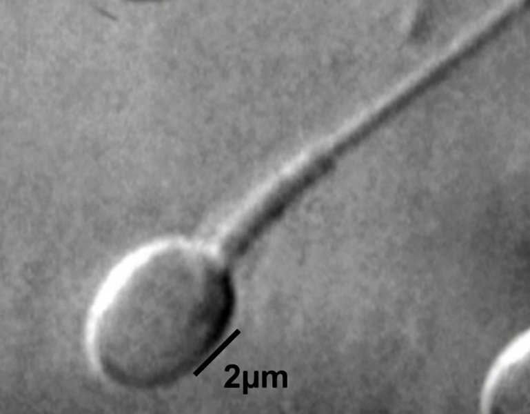
Single Human Spermatozoa Morphology
A: Normal spermatozoa observed at high magnification (≥ 8400×, in original figure)
A spermatozoon was classified as morphologically normal when it exhibited a normal nucleus (smooth, symmetric and oval nucleus, width 3.28 +/- 0.20 micron, length 4.75 +/- 0.20 micron/absence of vacuoles occupying > 4% of nuclear area) as well as acrosome, post-acrosomal lamina, neck and tail, besides not presenting cytoplasm around the head.
- Spermatozoa Images: Spermatozoa BF | Spermatozoon BF | Spermatozoon EM | Spermatozoon EM | Historic drawing | Category:Spermatozoa | Spermatozoa Development | Testis Development
Reference
<pubmed>20529256</pubmed>| Reprod Biol Endocrinol.
Copyright
© 2010 Oliveira et al; licensee BioMed Central Ltd. This is an Open Access article distributed under the terms of the Creative Commons Attribution License (http://creativecommons.org/licenses/by/2.0), which permits unrestricted use, distribution, and reproduction in any medium, provided the original work is properly cited.
Original File name: Figure 1. http://www.rbej.com/content/8/1/56/figure/F1 extracted from original figure, cropped, despeckeled and size adjusted.
File history
Click on a date/time to view the file as it appeared at that time.
| Date/Time | Thumbnail | Dimensions | User | Comment | |
|---|---|---|---|---|---|
| current | 10:51, 22 September 2010 |  | 1,000 × 780 (53 KB) | S8600021 (talk | contribs) | ==Single Human Spermatozoa Morphology== A: Normal spermatozoa observed at high magnification (≥ 8400×) A spermatozoon was classified as morphologically normal when it exhibited a normal nucleus (smooth, symmetric and oval nucleus, width 3.28 +/- 0.20 |
You cannot overwrite this file.
File usage
The following page uses this file: