File:Sickle cell RBC SEM01.jpg

Original file (1,000 × 750 pixels, file size: 219 KB, MIME type: image/jpeg)
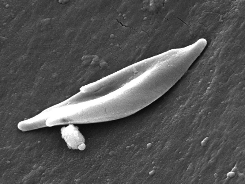
Human sickle cell RBC scanning electron micrograph (SEM)
This scanning electron micrograph (SEM) shows the ultrastructural morphology of a sickle cell RBC found in a blood specimen of an 18 year old female patient with sickle cell anemia, (HbSS).
People who have this form of sickle cell disease inherit two sickle cell genes (“S”), one from each parent. This is commonly called “sickle cell anemia”, and is usually the most severe form of the disease.
The name comes from the "sickle" shape of the RBC compared to the normal "donut" shape.
- Links: sickle cell disease | blood | cardiovascular abnormalities
Reference
CDC/ Sickle Cell Foundation of Georgia: Jackie George, Beverly Sinclair (2009)
http://phil.cdc.gov/phil/details.asp?pid=11687
Copyright
This image is in the public domain and thus free of any copyright restrictions. As a matter of courtesy we request that the content provider be credited and notified in any public or private usage of this image.
Cite this page: Hill, M.A. (2024, April 25) Embryology Sickle cell RBC SEM01.jpg. Retrieved from https://embryology.med.unsw.edu.au/embryology/index.php/File:Sickle_cell_RBC_SEM01.jpg
- © Dr Mark Hill 2024, UNSW Embryology ISBN: 978 0 7334 2609 4 - UNSW CRICOS Provider Code No. 00098G
File history
Click on a date/time to view the file as it appeared at that time.
| Date/Time | Thumbnail | Dimensions | User | Comment | |
|---|---|---|---|---|---|
| current | 04:22, 15 June 2015 |  | 1,000 × 750 (219 KB) | Z8600021 (talk | contribs) | 11687-sickle-cell-RBC.jpg |
You cannot overwrite this file.
File usage
The following 2 pages use this file: