File:Sea urchin Wnt patterning model.jpg

Original file (1,029 × 1,000 pixels, file size: 110 KB, MIME type: image/jpeg)
Sea urchin Wnt patterning model
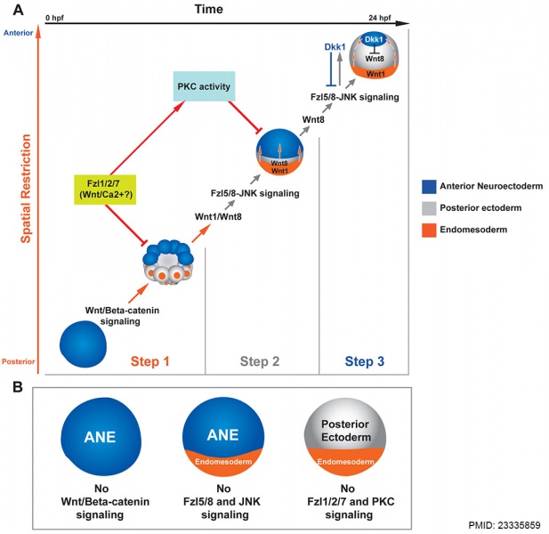
Three-step model for the balance of Wnt signaling interactions during anterior neuroectoderm (ANE) restriction.
(A) The y-axis monitors the progress of spatial restriction of the ANE (blue), and the x-axis indicates the developmental timing of ANE restriction. Each image represents the position of the ANE in time and space.
(Step 1) Initially, the maternal regulatory state in the absence of Wnt signaling supports ANE specification throughout the embryo. Then, nβ-catenin signaling in the posterior half of the embryo activates an unknown negative regulatory activity that blocks the accumulation of ANE factors, either by blocking their transcription directly or the activity of their ubiquitously expressed maternal activators.
(Step 2) As development progresses, posterior nβ-catenin activates production of at least two Wnt ligands, Wnt1 and Wnt8, that are necessary to initiate the ANE restriction mechanism in posterior ectoderm beginning at the 60-cell stage. These secreted ligands signal through the Wnt receptor, Fzl5/8, activating the JNK pathway. The Wnt/JNK pathway progressively down-regulates expression of ANE factors during early blastula stages in all but the most anterior ectoderm. During Step 1 and possibly Step 2, Fzl1/2/7 signaling attenuates the nβ-catenin- and Fzl5/8-JNK-mediated down-regulation of ANE factors, preventing complete shutdown of ANE specification. PKC activity also antagonizes Fzl5/8-JNK-mediated ANE restriction, downstream of Fzl1/2/7 signaling.
(Step 3) Expression of Wnt8, Wnt1, and/or another ligand X is activated in more anterior blastomeres. These ligands continue Fzl5/8-mediated ANE restriction until the late blastula-stage embryo activates production of the secreted Wnt antagonist, Dkk1, via Fzl5/8. Through negative feedback, Dkk1 limits Fzl5/8 activity, thereby defining the borders of the ANE. Orange arrows indicate the Wnt/β-catenin-mediated mechanism, gray arrows indicate the Wnt/JNK mediated mechanism, and red indicates Fzl1/2/7 and PKC interactions.
(B) Diagram showing the state of the ANE in embryos lacking either Wnt/β-catenin, Fzl5/8 and pJNK, or Fzl1/2/7 and pPKC signaling.
Reference
<pubmed>23335859</pubmed>| PLoS Biol.
Copyright
This is an open-access article, free of all copyright, and may be freely reproduced, distributed, transmitted, modified, built upon, or otherwise used by anyone for any lawful purpose. The work is made available under the Creative Commons CC0 public domain dedication.
Figure 7. Journal.pbio.1001467.g007.jpg doi:10.1371/journal.pbio.1001467.g007
File history
Click on a date/time to view the file as it appeared at that time.
| Date/Time | Thumbnail | Dimensions | User | Comment | |
|---|---|---|---|---|---|
| current | 18:04, 13 February 2014 |  | 1,029 × 1,000 (110 KB) | Z8600021 (talk | contribs) | ==Sea urchin Wnt patterning model== Three-step model for the balance of Wnt signaling interactions during ANE restriction. (A) The y-axis monitors the progress of spatial restriction of the ANE (blue), and the x-axis indicates the developmental timin... |
You cannot overwrite this file.
File usage
The following page uses this file: