File:Respiratory histology 10.jpg
From Embryology
Respiratory_histology_10.jpg (450 × 600 pixels, file size: 82 KB, MIME type: image/jpeg)
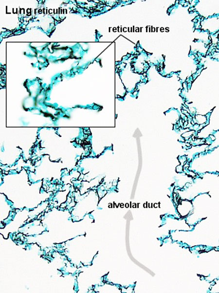
Lung Reticular Fibres
- Reticular and elastic fibres form the bulk of the connective tissue present in the walls of the alveoli.
- Collagenous fibres are sparse and fine in the alveolar walls.
- Note also that the tissue stained for reticular fibres looks much denser than the other sections. This lung collapsed prior to fixation because of the recoil of the elastic fibres.
Reticular Fibres - collagen type III
- Respiratory Histology: Bronchiole | Alveolar Duct | Alveoli | EM Alveoli septum | Alveoli Elastin | Trachea 1 | Trachea 2 | labeled lung | unlabeled lung | Respiratory Bronchiole | Lung Reticular Fibres | Nasal Inferior Concha | Nasal Respiratory Epithelium | Olfactory Region overview | Olfactory Region Epithelium | Histology Stains
File history
Click on a date/time to view the file as it appeared at that time.
| Date/Time | Thumbnail | Dimensions | User | Comment | |
|---|---|---|---|---|---|
| current | 22:51, 28 February 2012 |  | 450 × 600 (82 KB) | Z8600021 (talk | contribs) | ==Lung Reticular Fibres== {{Respiratory Histology}} |
You cannot overwrite this file.
