File:Placenta histology 005.jpg

Original file (1,280 × 960 pixels, file size: 433 KB, MIME type: image/jpeg)
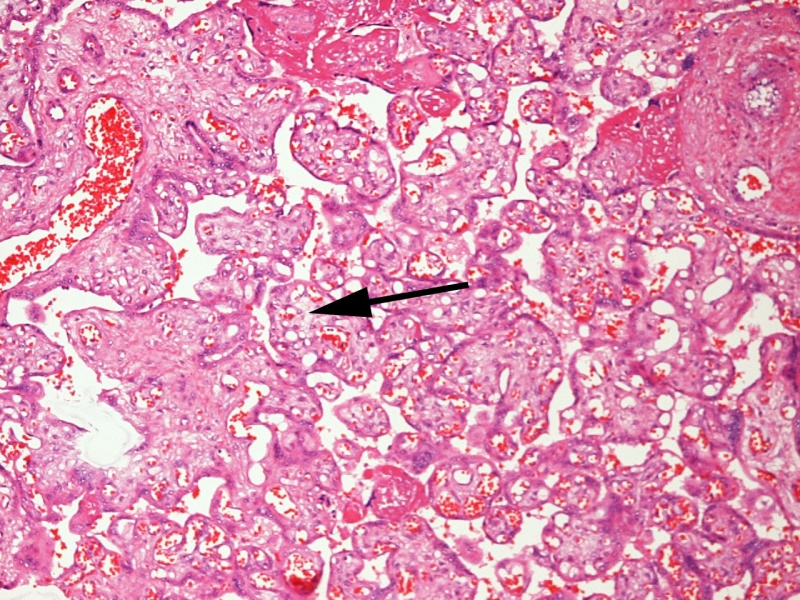
Placenta Histology - Chorangiosis with Gastroschisis
A 10× photomicrograph of chorangiosis in a placenta from a woman who delivered a patient with gastroschisis. The arrow points to an area with multiple vascular channels. Diffuse chorangiosis was defined as ≥ 10 capillaries in ≥ 10 terminal villi in 10 fields at 10× magnification in each of 3 areas (slides). Red blood cells can be seen in many of the capillaries. Capillary proliferation can be seen in numerous terminal villi.
References
<pubmed> 22004141 </pubmed>| BMC Pediatr.
Copyright
© 2011 Payne et al; licensee BioMed Central Ltd.
This is an Open Access article distributed under the terms of the Creative Commons Attribution License (http://creativecommons.org/licenses/by/2.0), which permits unrestricted use, distribution, and reproduction in any medium, provided the original work is properly cited. Payne et al. BMC Pediatrics 2011 11:90 doi:10.1186/1471-2431-11-90
Figure 6. http://www.biomedcentral.com/1471-2431/11/90/figure/F6
Cite this page: Hill, M.A. (2024, April 22) Embryology Placenta histology 005.jpg. Retrieved from https://embryology.med.unsw.edu.au/embryology/index.php/File:Placenta_histology_005.jpg
- © Dr Mark Hill 2024, UNSW Embryology ISBN: 978 0 7334 2609 4 - UNSW CRICOS Provider Code No. 00098G
File history
Click on a date/time to view the file as it appeared at that time.
| Date/Time | Thumbnail | Dimensions | User | Comment | |
|---|---|---|---|---|---|
| current | 11:24, 23 May 2012 |  | 1,280 × 960 (433 KB) | Z8600021 (talk | contribs) | ==Placenta Chorangiosis with Gastroschisis== |
You cannot overwrite this file.
File usage
The following page uses this file: