File:Periosteum.jpg
From Embryology
No higher resolution available.
Periosteum.jpg (500 × 333 pixels, file size: 34 KB, MIME type: image/jpeg)
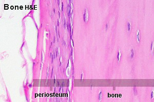
Bone Periosteum
- Bone is surrounded by a layer of dense connective tissue, the periosteum.
- A thin layer of cell-rich connective tissue, the endosteum, lines the surface of the bone facing the marrow cavity.
- Both the periosteum and the endosteum possess osteogenic potency.
- Following injury, cells in these layers may differentiate into osteoblasts (bone forming cells) which become involved in the repair of damage to the bone.
Links: Histology | Histology Stains | Blue Histology images copyright Lutz Slomianka 1998-2009. The literary and artistic works on the original Blue Histology website may be reproduced, adapted, published and distributed for non-commercial purposes. See also the page Histology Stains.
Cite this page: Hill, M.A. (2024, April 20) Embryology Periosteum.jpg. Retrieved from https://embryology.med.unsw.edu.au/embryology/index.php/File:Periosteum.jpg
- © Dr Mark Hill 2024, UNSW Embryology ISBN: 978 0 7334 2609 4 - UNSW CRICOS Provider Code No. 00098G
Original file name: Pos20he.jpg
File history
Click on a date/time to view the file as it appeared at that time.
| Date/Time | Thumbnail | Dimensions | User | Comment | |
|---|---|---|---|---|---|
| current | 14:40, 18 February 2013 | | 500 × 333 (34 KB) | Z8600021 (talk | contribs) | Increase image size and adjust contrast. |
| 11:26, 11 September 2009 |  | 300 × 200 (18 KB) | S8600021 (talk | contribs) | Bone is surrounded by a layer of dense connective tissue, the periosteum. A thin layer of cell-rich connective tissue, the endosteum, lines the surface of the bone facing the marrow cavity. Both the periosteum and the endosteum possess osteogenic potenc |
You cannot overwrite this file.