File:Paramesonephric duct.jpg
From Embryology
No higher resolution available.
Paramesonephric_duct.jpg (423 × 478 pixels, file size: 40 KB, MIME type: image/jpeg)
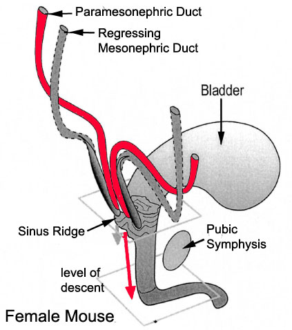
Paramesonephric Duct
This mouse image shows the relationship between the mesonephric and paramesonephric ducts opening into the urogenital sinus.
- The paramesonephric duct began as an infold of surface epithelium lying along the surface of the genital ridge.
- Estrogens, both maternal and fetal, stimulate its development and that eventually of the external female fetal genital structures.
- In contrast, the mesonephric duct regresses, remnants of this duct may remain lying within the broad ligament.
(Image modified from Biol Reprod. 2002 Oct;67(4):1353-9. PMID: 12297555)
Reference
<pubmed>12297555</pubmed>
File history
Click on a date/time to view the file as it appeared at that time.
| Date/Time | Thumbnail | Dimensions | User | Comment | |
|---|---|---|---|---|---|
| current | 22:49, 21 September 2009 | | 423 × 478 (40 KB) | S8600021 (talk | contribs) |
You cannot overwrite this file.
File usage
The following 13 pages use this file:
- 2009 Lecture 16
- 2010 Lecture 16
- 2011 Lab 8 - Fetal
- 2011 Lecture 16
- 2014 Group Project 4
- ANAT2341 Lab 8 - Fetal
- BGDB Sexual Differentiation - Fetal
- BGD Lecture - Sexual Differentiation
- Lecture - Genital Development
- REI - Reproductive Medicine Seminar 2018
- Royal Hospital for Women - Reproductive Medicine Seminar 2018
- Uterus Development
- Talk:2014 Group Project 4