File:Oxytocin receptor pathways.jpg
Oxytocin_receptor_pathways.jpg (720 × 512 pixels, file size: 88 KB, MIME type: image/jpeg)
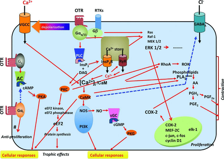
Oxytocin Receptor Pathways
| Oxytocin receptor (OTR) activation leads to three different GTP-binding protein mechanisms.
The major mechanism is mediated by the Gq/PLC/InsP3 pathway. When oxytocin binds to OTR, it activates Gαq/11 and then phospholipase C (PLC), which induces the cleavage of PIP2 to inositoltrisphosphate (InsP3) and diacylglycerol (DAG). InsP3 induces Ca2+ release from Ca2+ stores via InsP3R and, in some cells, causes Ca2+-induced Ca2+ release (CICR) via the ryanodine receptor (RyR). The activation of Gq also causes membrane depolarization*, which, in turn, activates VGCCs and then facilitates Ca2+ entry through VGCCs. Thus, increased cytosolic Ca2+ ([Ca2+]i) stimulates CaMK after binding to the Ca2+ binding protein Calmodulin. The Ca2+/CaM complex then activates CaMK and causes various cellular responses, such as smooth muscle contractions, or induces the activation of several different types of enzymes, such as NOS or PI3K. DAG causes protein kinase C (PKC) activation and also various cellular responses. Additional pathways activated through the OTR include the MAP-kinase (MAPK) and the Rho kinase pathways. The increased transcription of COX2 mediates the increased production and secretion of prostaglandins. The OTR-mediated opening of Ca2+ channels is likely mediated through free Gβγ subunits. The OT receptor is known to be coupled with the other G proteins, Gs and Gi, both of which are linked with the AC pathway. The proliferative effects involve MAPK-mediated activation of specific gene transcription. The trophic effects are mediated via a PKC-mediated activation of eEF2. Activation of the Rho and MAP kinase pathways, the increase in intracellular Ca2+ and the increased prostaglandin secretion all contribute to the contractile effects. The antiproliferative effects observed in certain cells types appear to be mediated via αi G protein subunits.
|
Legend:
|
| About Oxytocin Hormone | |
|---|---|
| Structure | Function |
|
|
| Links: Birth | Hypothalamus | Endocrine | Mammary Gland | Milk | image - Oxytocin receptor pathways | |
- Links: Birth | Endocrine System Development
Reference
Viero C, Shibuya I, Kitamura N, Verkhratsky A, Fujihara H, Katoh A, Ueta Y, Zingg HH, Chvatal A, Sykova E & Dayanithi G. (2010). REVIEW: Oxytocin: Crossing the bridge between basic science and pharmacotherapy. CNS Neurosci Ther , 16, e138-56. PMID: 20626426 DOI.
Copyright
Re-use of this article is permitted in accordance with the Terms and Conditions set out at http://www3.interscience.wiley.com/authorresources/onlineopen.html
Re-use of this article is permitted in accordance with the Creative Commons Deed, Attribution 2.5, which does not permit commercial exploitation.
From: CNS Neurosci Ther. 2010 October; 16(5): e138–e156. doi: 10.1111/j.1755-5949.2010.00185.x.
Original file name: Figure 2 Cns0016-e138-f2.jpg http://www.ncbi.nlm.nih.gov/pmc/articles/PMC2972642/figure/fig02/
Cite this page: Hill, M.A. (2024, April 19) Embryology Oxytocin receptor pathways.jpg. Retrieved from https://embryology.med.unsw.edu.au/embryology/index.php/File:Oxytocin_receptor_pathways.jpg
- © Dr Mark Hill 2024, UNSW Embryology ISBN: 978 0 7334 2609 4 - UNSW CRICOS Provider Code No. 00098G
File history
Click on a date/time to view the file as it appeared at that time.
| Date/Time | Thumbnail | Dimensions | User | Comment | |
|---|---|---|---|---|---|
| current | 12:44, 20 December 2010 | | 720 × 512 (88 KB) | S8600021 (talk | contribs) | ==Oxytocin Receptor Pathways== Oxytocin receptor (OTR) activation leads to three different GTP-binding protein mechanisms. The major mechanism is mediated by the Gq/PLC/InsP3 pathway. When oxytocin binds to OTR, it activates Gαq/11 and then phospholip |
You cannot overwrite this file.
File usage
The following 2 pages use this file: