File:Ovary histology 001.jpg
Original file (1,280 × 1,024 pixels, file size: 360 KB, MIME type: image/jpeg)
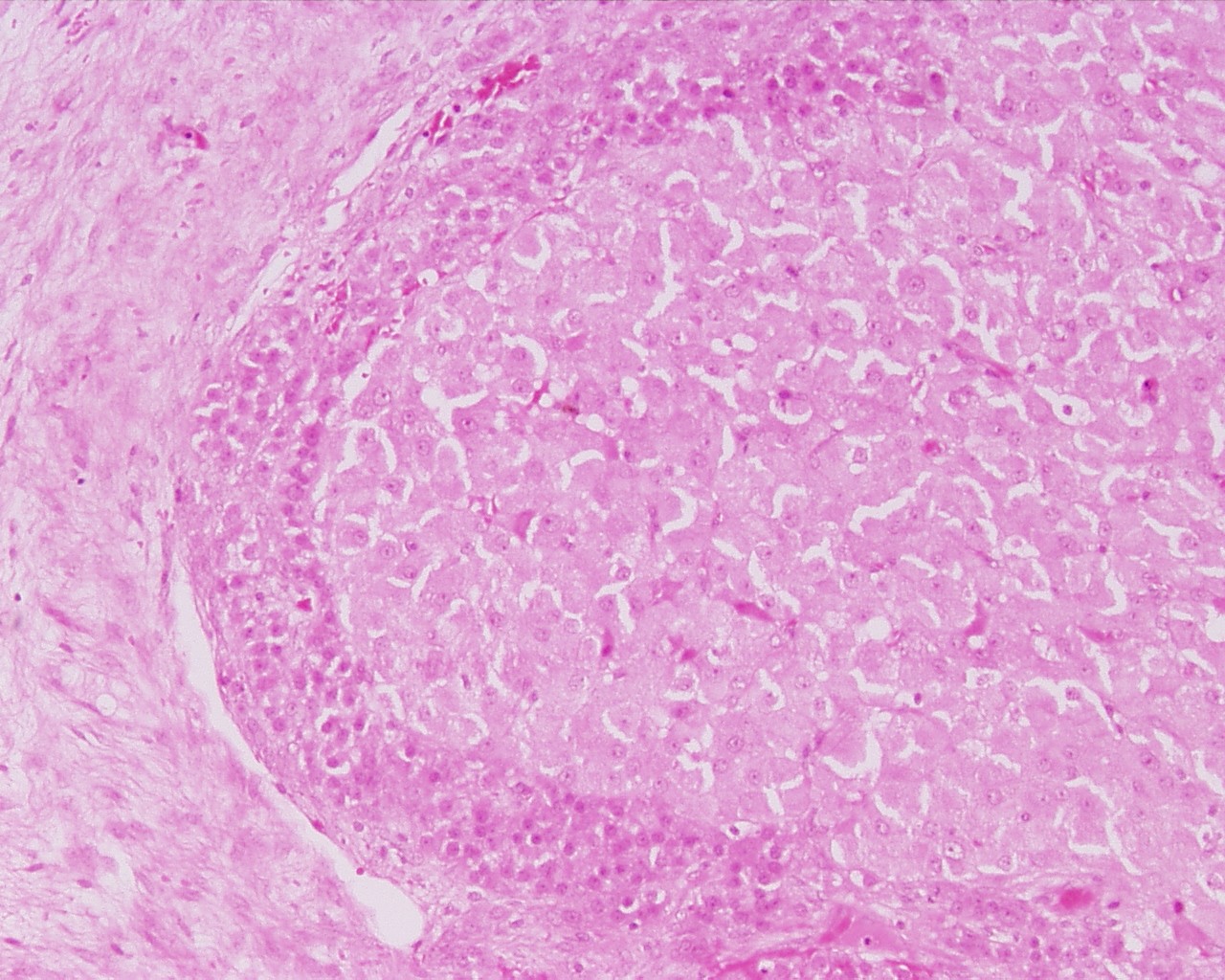
Ovary - Corpus Luteum
The corpus luteum is formed by both granulosa cells and thecal cells after ovulation has occurred. The wall of the follicle collapses into a folded structure, which is characteristic for the corpus luteum. Vascularization increases and a connective tissue network is formed. Theca interna cells and granulosa cells triple in size and start accumulating lutein (Which hormone stimulates this process? Where is this hormone produced?) within a few hours after ovulation. They are now called granulosa lutein cells and theca lutein cells and produce progesterone and oestrogens.
Hormone secretion in the corpus luteum ceases within 14 days after ovulation if the oocyte is not fertilised. In this case, the corpus luteum degenerates into a corpus albicans - whitish scar tissue within the ovaries.
Hormone secretion continues for 2-3 month after ovulation if fertilisation occurs.
The dark cell represent theca lutein cell the lighter ones are granulosa lutein cells.
Ovary histology: Tunica Albuginea x20 | Tunica albuginea, Germinal epithelium x40 |
Primary follicle, primordial follicle, oocyte, x40 | Secondary follicle, cumulus oophorus, zona pelucida, granulosa cells, oocyte x20 | Corpus luteum, theca lutein cells, granulosa lutein cells, Loupe | Corpus luteum, theca lutein cells, granulosa lutein cells, x10 | Corpus luteum, theca lutein cells, granulosa lutein cells, x40 | Corpus albicans, primary follicle, primordial follicle, granulosa cells, oocyte x20 | Menstrual Cycle | Ovary Development
Links: Histology | Histology Stains | Blue Histology images copyright Lutz Slomianka 1998-2009. The literary and artistic works on the original Blue Histology website may be reproduced, adapted, published and distributed for non-commercial purposes. See also the page Histology Stains.
Cite this page: Hill, M.A. (2024, April 18) Embryology Ovary histology 001.jpg. Retrieved from https://embryology.med.unsw.edu.au/embryology/index.php/File:Ovary_histology_001.jpg
- © Dr Mark Hill 2024, UNSW Embryology ISBN: 978 0 7334 2609 4 - UNSW CRICOS Provider Code No. 00098G
Corpus luteum, H&E theca lutein cells, granulosa lutein cells, x10
File history
Click on a date/time to view the file as it appeared at that time.
| Date/Time | Thumbnail | Dimensions | User | Comment | |
|---|---|---|---|---|---|
| current | 21:07, 23 February 2011 | | 1,280 × 1,024 (360 KB) | S8600021 (talk | contribs) | File:Ovary_histology_001.jpg Corpus_luteum_H&E_reproductive_system,_female,_theca_lutein_cells,_granulosa_lutein_cellsx10.jpg {{Ovary Histology}} {{Template:Blue Histology}} Category:Genital Category:Histology Category:Ovary [[Category:O |
You cannot overwrite this file.
File usage
The following 5 pages use this file: