File:Mouse renal ureteropelvic junction obstruction 01.jpg

Original file (1,200 × 1,243 pixels, file size: 332 KB, MIME type: image/jpeg)
Mouse Renal Ureteropelvic Junction Obstruction
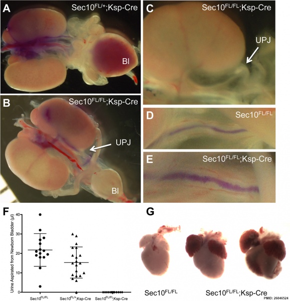
Hydronephrosis in Sec10FL/FL;Ksp-Cre kidneys is due to physical obstructions in ureters at the UPJ, resulting in anuria and heart failure.
- A, B - Urinary tracts, including both kidneys, intact ureters, and bladder were removed from mutant Sec10FL/FL;Ksp-Cre mice and littermate controls (Sec10FL/FL and Sec10Fl/+;Ksp-Cre) at E18.5 and P0. Blue dye was injected into the renal pelvis, and in control kidneys the dye migrated down the ureters and accumulated in the bladder as expected (representative E18.5 sample shown in A). In every Sec10FL/FL;Ksp-Cre kidney with hydronephrosis tested at E18.5 and P0, the dye stopped at the UPJ (arrow, representative E18.5 sample shown in B).
- C - Occasionally in the dissected mutant newborn kidneys with hydronephrosis, a microscopic examination revealed a deposit of white debris within the ureter above the UPJ region, also suggesting a physical blockage of the ureter lumen (arrow, C).
- D, E - In one of the few newborn Sec10FL/FL;Ksp-Cre kidneys that did not show hydronephrosis, dye injections traveled to the bladder, but revealed a visibly abnormal ureter lumen with rough irregular edges (E) not observed in controls (D).
- F - Aspirations from bladders of newborn pups confirmed that no urine was present in the bladders of Sec10FL/FL;Ksp-Cre pups with bilateral hydronephrosis, compared with a normal distribution found in littermate controls (shown are means ± SD).
- G - All newborn Sec10FL/FL;Ksp-Cre pups with bilateral obstructions and hydronephrosis died 6–14 hours after birth, with necropsies revealing heart wall distension and cardiac hemorrhaging. Shown are two representative Sec10FL/FL;Ksp-Cre hearts (right) dissected immediately after death (12-hours post-birth), compared to an age-matched littermate control heart (left).
SEC10 is a member of the exocyst complex, which was identified as essential for exocytosis in yeast. SEC10L1 has been suggested as a component of the human exocyst complex (octameric protein complex) essential for post-Golgi traffic.
Reference
<pubmed>26046524</pubmed>| PLoS One.
Fogelgren B, Polgar N, Lui VH, Lee AJ, Tamashiro K-KA, Napoli JA, et al. (2015) Urothelial Defects from Targeted Inactivation of Exocyst Sec10 in Mice Cause Ureteropelvic Junction Obstructions. PLoS ONE 10(6): e0129346. doi:10.1371/journal.pone.0129346
Copyright
© 2015 Fogelgren et al. This is an open access article distributed under the terms of the Creative Commons Attribution License, which permits unrestricted use, distribution, and reproduction in any medium, provided the original author and source are credited
Fig 3. journal.pone.0129346.g003.jpg doi:10.1371/journal.pone.0129346.g003
File history
Click on a date/time to view the file as it appeared at that time.
| Date/Time | Thumbnail | Dimensions | User | Comment | |
|---|---|---|---|---|---|
| current | 05:34, 7 June 2015 | | 1,200 × 1,243 (332 KB) | Z8600021 (talk | contribs) | Mouse ureteropelvic junction obstruction 01.jpg Fig 3. Hydronephrosis in Sec10FL/FL;Ksp-Cre kidneys is due to physical obstructions in ureters at the UPJ, resulting in anuria and heart failure. (A, B) Urinary tracts, including both kidneys, intact u... |
You cannot overwrite this file.
File usage
The following page uses this file: