File:Mouse placenta 02.jpg
Mouse_placenta_02.jpg (464 × 220 pixels, file size: 21 KB, MIME type: image/jpeg)
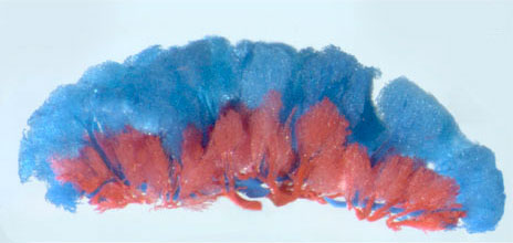
Mouse placental blood vessels
Mouse (E16.5) lateral view (maternal side at bottom)
- Venous side - red resin
- Arterial side - blue resin
Resin casts are generated by filling the existing vascular beds (day 16.5 p.c.) with a resin that sets and the surrounding materials are then removed, leaving just the vascular beds.
- Links: Mouse placental blood vessels Superior View | Mouse placental blood vessels lateral View | [Mouse Development]] | Placenta Development
Thanks to Dr Sally Dunwoodie, Developmental Biology Program, Victor Chang Cardiac Research Institute who provided both images and information on (mouse) placental development. (Email: Sally Dunwoodie)
Reference
Citation: Cited1 is required in trophoblasts for placental development and for embryo growth and survival. Rodriguez TA, Sparrow DB, Scott AN, Withington SL, Preis JI, Michalicek J, Clements M, Tsang TE, Shioda T, Beddington RS, Dunwoodie SL. Mol Cell Biol. 2004 Jan;24(1):228-44. PMID: 14673158
- "Cited1 is a transcriptional cofactor that interacts with Smad4, estrogen receptors alpha and beta, TFAP2, and CBP/p300. It is expressed in a restricted manner in the embryo as well as in extraembryonic tissues during embryonic development. In this study we report the engineering of a loss-of-function Cited1 mutation in the mouse. Cited1 null mutants show growth restriction at 18.5 days postcoitum, and most of them die shortly after birth. Half the heterozygous females, i.e., those that carry a paternally inherited wild-type Cited1 allele, are similarly affected. Cited1 is normally expressed in trophectoderm-derived c ells of the placenta; however, in these heterozygous females, Cited1 is not expressed in these cells. This occurs because Cited1 is located on the X chromosome, and thus the wild-type Cited1 allele is not expressed because the paternal X chromosome is preferentially inactivated. Loss of Cited1 resulted in abnormal placental development. In mutants, the spongiotrophoblast layer is irregular in shape and enlarged while the labyrinthine layer is reduced in size. In addition, the blood spaces within the labyrinthine layer are disrupted; the maternal sinusoids are considerably larger in mutants, leading to a reduction in the surface area available for nutrient exchange. We conclude that Cited1 is required in trophoblasts for normal placental development and subsequently for embryo viability."
File history
Click on a date/time to view the file as it appeared at that time.
| Date/Time | Thumbnail | Dimensions | User | Comment | |
|---|---|---|---|---|---|
| current | 11:10, 16 April 2010 |  | 464 × 220 (21 KB) | S8600021 (talk | contribs) | '''Mouse placental blood vessels''' (E16.5) lateral view (maternal side at bottom) * Venous side - red resin * Arterial side - blue resin Resin casts are generated by filling the existing vascular beds (day 16.5 p.c.) with a resin that sets and the surr |
You cannot overwrite this file.
File usage
The following 5 pages use this file:
