File:Mouse-axial rotation.jpg

Original file (957 × 808 pixels, file size: 87 KB, MIME type: image/jpeg)
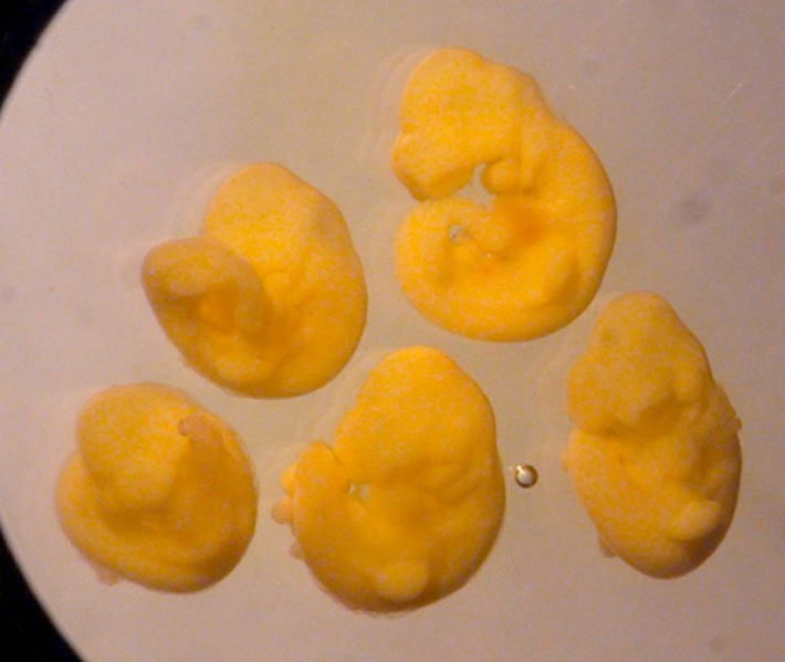
Litter of 5 fetuses of which two show clockwise rather than counter-clockwise axial rotation.
A litter of five fetuses from 8-cell stage E reconstructions that have completed axial rotation and shown with their left side oriented uppermost. The tail is curled to the left of the trunk in the upper two and to the right in lower three.
Figure 3. pone.0009610.g003.jpg
Normal Bias in the Direction of Fetal Rotation Depends on Blastomere Composition during Early Cleavage in the Mouse Richard L. Gardner PLoS One. 2010; 5(3): e9610. Published online 2010 March 10. doi: 10.1371/journal.pone.0009610. PMCID: PMC2835742 | PLoS
PLoS One. 2010; 5(3): e9610. Published online 2010 March 10. doi: 10.1371/journal.pone.0009610.
Copyright Richard L. Gardner. This is an open-access article distributed under the terms of the Creative Commons Attribution License, which permits unrestricted use, distribution, and reproduction in any medium, provided the original author and source are credited.
File history
Click on a date/time to view the file as it appeared at that time.
| Date/Time | Thumbnail | Dimensions | User | Comment | |
|---|---|---|---|---|---|
| current | 08:46, 5 April 2010 |  | 957 × 808 (87 KB) | S8600021 (talk | contribs) | Litter of 5 fetuses of which two show clockwise rather than counter-clockwise axial rotation. A litter of five fetuses from 8-cell stage E reconstructions that have completed axial rotation and shown with their left side oriented uppermost. The tail is c |
You cannot overwrite this file.
File usage
The following page uses this file: