File:Mouse- tooth histology.jpg
Mouse-_tooth_histology.jpg (500 × 503 pixels, file size: 42 KB, MIME type: image/jpeg)
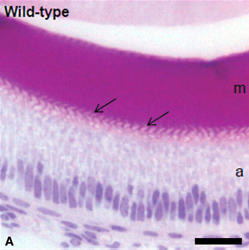
Mouse developing tooth histology
acrylic resin histology
(A) Semi-thin, transverse sections of the secretory zone of WT mice revealed pale-staining ameloblasts and an intensely eosinophilic, enamel matrix. The interdigitations of the ameloblast Tomes’ processes were clearly evident in the region where new enamel matrix was forming (arrows).
Legend
- m - enamel matrix
- a - ameloblasts
- n - nuclei
Scale bar 25 µm
Original File Name: Figure 6. http://hmg.oxfordjournals.org/content/vol19/issue7/images/large/ddq00106.jpeg (Extract from full figure)
Reference
<pubmed>20067920</pubmed>
© The Author 2010. Published by Oxford University Press
This is an Open Access article distributed under the terms of the Creative Commons Attribution Non-Commercial License (http://creativecommons.org/licenses/by-nc/2.5/uk) which permits unrestricted non-commercial use, distribution, and reproduction in any medium, provided the original work is properly cited.
File history
Click on a date/time to view the file as it appeared at that time.
| Date/Time | Thumbnail | Dimensions | User | Comment | |
|---|---|---|---|---|---|
| current | 12:37, 22 April 2010 |  | 500 × 503 (42 KB) | S8600021 (talk | contribs) | Mouse developing tooth acrylic resin histology. (A) Semi-thin, transverse sections of the secretory zone of WT mice revealed pale-staining ameloblasts and an intensely eosinophilic, enamel matrix. The interdigitations of the ameloblast Tomes’ processe |
You cannot overwrite this file.
File usage
The following page uses this file:
