File:Monkey- ovary primordial follicle.jpg

Original file (1,000 × 800 pixels, file size: 292 KB, MIME type: image/jpeg)
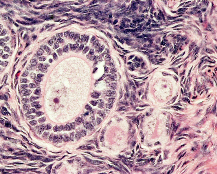
Ovary Histology
Ovary, monkey (Stain - Haematoxylin Eosin)
The large follicle in the centre of the image contains an oocyte surrounded by a double layer of granulosa cells, outside of this layer is the thecae layer.
- Links: Ovary Development
Links: Histology | Histology Stains | Blue Histology images copyright Lutz Slomianka 1998-2009. The literary and artistic works on the original Blue Histology website may be reproduced, adapted, published and distributed for non-commercial purposes. See also the page Histology Stains.
Cite this page: Hill, M.A. (2024, April 19) Embryology Monkey- ovary primordial follicle.jpg. Retrieved from https://embryology.med.unsw.edu.au/embryology/index.php/File:Monkey-_ovary_primordial_follicle.jpg
- © Dr Mark Hill 2024, UNSW Embryology ISBN: 978 0 7334 2609 4 - UNSW CRICOS Provider Code No. 00098G
File history
Click on a date/time to view the file as it appeared at that time.
| Date/Time | Thumbnail | Dimensions | User | Comment | |
|---|---|---|---|---|---|
| current | 21:57, 30 May 2010 | | 1,000 × 800 (292 KB) | S8600021 (talk | contribs) | |
| 23:13, 21 April 2010 |  | 1,280 × 1,024 (427 KB) | S8600021 (talk | contribs) | Ovary Histology ovary, monkey H&E reproductive system, female, primary follicle, primordial follicle, oocyte Histology image H&E high power Image Source: Lutz Slomianka, UWA Blue Histology Ova41he.jpg http://www.lab.anhb.uwa.edu.au/mb140/CorePages/ |
You cannot overwrite this file.
File usage
The following 2 pages use this file: