File:Model of human pancreatic islet.jpg
Original file (482 × 702 pixels, file size: 237 KB, MIME type: image/jpeg)
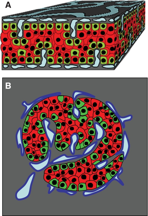
Model of endocrine cell and vessel organization in human islets
A α-Cells (green) and β-cells (red) are organized into a thick folded plate lined at both sides with vessels (blue).
- α-Cells are mostly at the periphery of the plate and in close contact with vessels.
- β-Cells occupy a more central part of the plate and most of them develop cytoplasmic extension that runs between α-cells and reaches the surface of vessels.
B The plate with adjacent vessels is folded so that it forms an islet.
Original file name: FIG. 7. Zdb0051061080007.jpg http://www.ncbi.nlm.nih.gov/pmc/articles/PMC2857900/figure/F7/
Reference
Bosco D, Armanet M, Morel P, Niclauss N, Sgroi A, Muller YD, Giovannoni L, Parnaud G & Berney T. (2010). Unique arrangement of alpha- and beta-cells in human islets of Langerhans. Diabetes , 59, 1202-10. PMID: 20185817 DOI.
Copyright
© 2010 by the American Diabetes Association
Readers may use this article as long as the work is properly cited, the use is educational and not for profit, and the work is not altered. See http://creativecommons.org/licenses/by-nc-nd/3.0/ for details.
Cite this page: Hill, M.A. (2024, April 23) Embryology Model of human pancreatic islet.jpg. Retrieved from https://embryology.med.unsw.edu.au/embryology/index.php/File:Model_of_human_pancreatic_islet.jpg
- © Dr Mark Hill 2024, UNSW Embryology ISBN: 978 0 7334 2609 4 - UNSW CRICOS Provider Code No. 00098G
File history
Click on a date/time to view the file as it appeared at that time.
| Date/Time | Thumbnail | Dimensions | User | Comment | |
|---|---|---|---|---|---|
| current | 13:30, 8 August 2011 | | 482 × 702 (237 KB) | S8600021 (talk | contribs) | ==Model of endocrine cell and vessel organization in human islets== A: α-Cells (green) and β-cells (red) are organized into a thick folded plate lined at both sides with vessels (blue). α-Cells are mostly at the periphery of the plate and in close con |
You cannot overwrite this file.
File usage
The following 2 pages use this file: