File:Limb changing 3D geometry.jpg

Original file (1,000 × 1,062 pixels, file size: 196 KB, MIME type: image/jpeg)
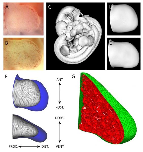
The changing 3D geometry during limb development
(A) The right hind limb bud at stage E11.0 from the freshly dissected embryo, and (B) a right hind limb bud from another embryo at stage E11.25, 6 h older than (A).
(C) The OPT scans were converted into an iso-surface, a 3D contour of the embryo.
The limb bud shapes St0, (D) and St1 (E) were virtually dissected from the whole-embryo iso-surface.
(F) A comparison between the two empirically measured shapes St0 (white) and St1 (blue) which highlights the shape change over 6 h of development. The main axes are shown anterio-posterior (AP), dorso-ventral (DV), and proximo-distal (PD).
(G) A virtual slice through the fully tetrahedtralised St0 mesh. The surface is discretised with a triangular mesh (green), and the internal mesenchymal volume is discretised with a 3D tetrahedral mesh (red).
Original file name: Figure 2. Journal.pbio.1000420.g002.png (resized using photoshop and converted to jpg)
Reference
<pubmed>20644711</pubmed>| PMC2903592 | PLoS
Citation: Boehm B, Westerberg H, Lesnicar-Pucko G, Raja S, Rautschka M, et al. (2010) The Role of Spatially Controlled Cell Proliferation in Limb Bud Morphogenesis. PLoS Biol 8(7): e1000420. doi:10.1371/journal.pbio.1000420
Copyright: © 2010 Boehm et al. This is an open-access article distributed under the terms of the Creative Commons Attribution License, which permits unrestricted use, distribution, and reproduction in any medium, provided the original author and source are credited.
File history
Click on a date/time to view the file as it appeared at that time.
| Date/Time | Thumbnail | Dimensions | User | Comment | |
|---|---|---|---|---|---|
| current | 10:21, 23 March 2011 |  | 1,000 × 1,062 (196 KB) | S8600021 (talk | contribs) | ==The changing 3D geometry during limb development== (A) The right hind limb bud at stage E11.0 from the freshly dissected embryo, and (B) a right hind limb bud from another embryo at stage E11.25, 6 h older than (A). (C) The OPT scans were converted i |
You cannot overwrite this file.
File usage
The following 2 pages use this file: