File:Keith1902 fig084.jpg

Original file (732 × 800 pixels, file size: 88 KB, MIME type: image/jpeg)
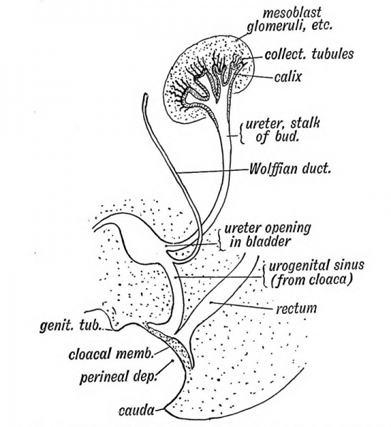
Fig. 84. The Termination of the Ureter in the Bladder and Sub-division of the Renal Bud
The connection of the stalk with the Wolffian duct is lost ; the termination of the ureter migrates along the duct until it reachei that part of the cloaca which afterwards forms the bladder (Fig 84). The dilated cephalic end of the bud divides into severa secondary buds. The dilated terminal part forms tb pelvis of the kidney, its infundibula and calyces. The tubule of the kidney are formed from groups of epithelial cells on the convex margin of the dilated end of the renal bud. They arise as tubular buds which grow out into the intermediate mesoblast glomeruli, etc. cell mass, subdividing as they grow spring out in groups, each group out forming inter at its mass,
| Historic Disclaimer - information about historic embryology pages |
|---|
 |
- The Uro-genital System: Fig. 79. Wolffian Body | Fig. 80. Wolffian and Genital Ridges | Fig. 81. Female Wolffian Body Remnants | Fig. 82. Male Wolffian Body Remnants |Fig. 83. Renal Bud | Fig. 84. Ureter in the Bladder | Fig. 85. Wolffian and Müllerian Ducts | Fig. 86. Genital Ducts 3rd month | Fig. 87. Müllerian Ducts 3rd month | Fig. 88. Uterus | Fig. 89. Uterus and Vagina | Fig. 90. Prostate remnants of Müllerian Ducts | Fig. 91. Prostate showing an unusual Uterus Masculinus | Fig. 92. Female Uro-genital Sinus | Fig. 93. Male Uro-genital Sinus | Fig. 94. Vagina and Uterus at 7th month | Fig. 95. Division of the Cloaca | Fig. 96. Imperforate Anus | Fig. 97. Cloacal Septum has failed to fuse with Perineal Septum | Fig. 98. The Uro-genital Cleft 2nd month | Fig. 99. Male bladder and urethra at birth | Fig. 100. Ectopia Vesicae | Fig. 101. Prostatic Tubules | Fig. 102. Testis in a foetus of 2.5 months | Fig. 103. Testis at the 6th month | Fig. 104. Inguinal Canal and Coverings of the Testis | Fig. 105. Processus Vaginalis | Figures
| Historic Disclaimer - information about historic embryology pages |
|---|
|
Human Embryology and Morphology (1902): Development or the Face | The Nasal Cavities and Olfactory Structures | Development of the Pharynx and Neck | Development of the Organ of Hearing | Development and Morphology of the Teeth | The Skin and its Appendages | The Development of the Ovum of the Foetus from the Ovum of the Mother | The Manner in which a Connection is Established between the Foetus and Uterus | The Uro-genital System | Formation of the Pubo-femoral Region, Pelvic Floor and Fascia | The Spinal Column and Back | The Segmentation of the Body | The Cranium | Development of the Structures concerned in the Sense of Sight | The Brain and Spinal Cord | Development of the Circulatory System | The Respiratory System | The Organs of Digestion | The Body Wall, Ribs, and Sternum | The Limbs | Figures | Embryology History
Reference
Keith A. Human Embryology and Morphology. (1902) London: Edward Arnold.
Cite this page: Hill, M.A. (2024, April 18) Embryology Keith1902 fig084.jpg. Retrieved from https://embryology.med.unsw.edu.au/embryology/index.php/File:Keith1902_fig084.jpg
- © Dr Mark Hill 2024, UNSW Embryology ISBN: 978 0 7334 2609 4 - UNSW CRICOS Provider Code No. 00098G
File history
Click on a date/time to view the file as it appeared at that time.
| Date/Time | Thumbnail | Dimensions | User | Comment | |
|---|---|---|---|---|---|
| current | 10:26, 7 January 2014 | | 732 × 800 (88 KB) | Z8600021 (talk | contribs) |
You cannot overwrite this file.
File usage
The following 4 pages use this file: