File:Keibel Mall 297.jpg
Keibel_Mall_297.jpg (371 × 291 pixels, file size: 16 KB, MIME type: image/jpeg)
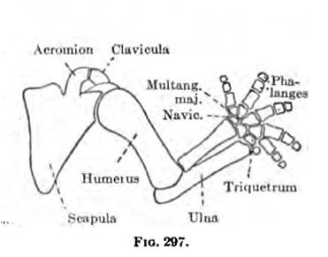
Fig. 297. Human Embryo Limb Skeleton
In an embryo 20 mm. long the cartilaginons anlages of various bones of the superior extremity are all well marked, except those of the distal row of the phalanges of the fingers. The clavicle extends from the acromion to the sternum. It is composed of a peculiar kind of precartitaginous tissue. The general shape of the other cartilages may be seen from Fig. 297. The spine of the scapula is not yet distinct. There are distinct coraccclavicular, costoclavicular, and interclavicular ligaments. There is no joint cavity at the shoulder, but a capsular and a coraeo-humeral ligament may be distinguished. The humerus has well-marked tuberosities and condyles. The ulna and radius are larger and longer than at the preceding stage. The olecranon, coraccid, and styloid processes are composed of cartilage and condensed tissue. The perichondrium about the ulna and radius is quite thick. The capsular and annular ligaments are present, but there are no joint cavities.
- Limb Images: 274-278 Spinal Column and Lower Limb | 279-284 Lower Limb | 285-288 Knee | 289 Os Coxae | 290 Femur | 291 Tibia | 292 Fibula | 293 Foot | 294 | 295 | 296 | 297 | 298-299 | 300 Forearm and Hand | 301 Upper Limb Joints | 302 Clavicle | Upper Limb Ossification 1 | Upper Limb Ossification 2 | Bone Development Timeline
- Skeleton and Connective Tissues: Connective Tissue Histogenesis | Skeletal Morphogenesis | Chorda Dorsalis | Vertebral Column and Thorax | Limb Skeleton | Skull Hyoid Bone Larynx
- KM Figure Links: The Germ Cells | Segmentation | First Primitive Segment | Gastrulation | External Form | Placenta | Axial Skeleton | Limb Skeleton | Skull | Muscular System
| Historic Disclaimer - information about historic embryology pages |
|---|
 |
Glossary Links
- Glossary: A | B | C | D | E | F | G | H | I | J | K | L | M | N | O | P | Q | R | S | T | U | V | W | X | Y | Z | Numbers | Symbols | Term Link
Cite this page: Hill, M.A. (2024, April 18) Embryology Keibel Mall 297.jpg. Retrieved from https://embryology.med.unsw.edu.au/embryology/index.php/File:Keibel_Mall_297.jpg
- © Dr Mark Hill 2024, UNSW Embryology ISBN: 978 0 7334 2609 4 - UNSW CRICOS Provider Code No. 00098G
File history
Click on a date/time to view the file as it appeared at that time.
| Date/Time | Thumbnail | Dimensions | User | Comment | |
|---|---|---|---|---|---|
| current | 18:51, 27 August 2012 | | 371 × 291 (16 KB) | Z8600021 (talk | contribs) | ==Fig. 297 Human Embryo Limb Skeleton== {{KM Skeleton}} {{Keibel_Mall Images}} |
You cannot overwrite this file.