File:Keibel1900 plate03.jpg
Original file (1,929 × 2,605 pixels, file size: 588 KB, MIME type: image/jpeg)
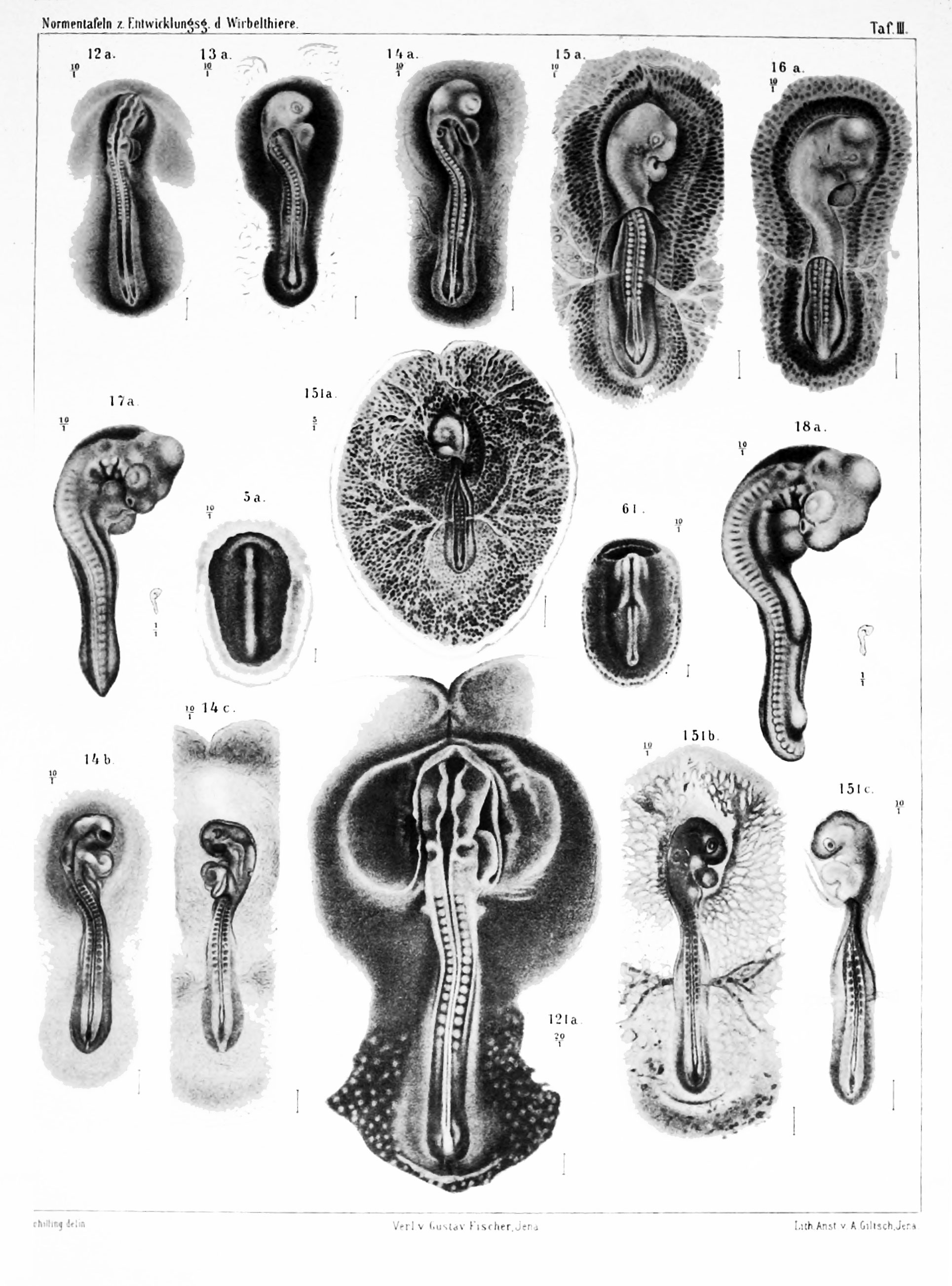
Plate 3
Figure legends text shown below is a slightly modified poor online translation. Original German text.
| Translation Request |
|---|
 If you would like to help with the online translation into English please contact me. |
| Historic Disclaimer - information about historic embryology pages |
|---|
|
Figure 12a
(S.N. 418; Tab. 18.)
The embryo, after the figures are 12 and 12a drawn, was a 46-h (1 day 22 hours) taken from incubated egg fixed with sublimate-acetic acid. The front end of the body has become pretty much lifted from the blastoderm and begins to rotate to the left side. The neural tube is closed except for a few cuts. It shows the three brain vesicles, the investments of the neuromeres are indicated, and the roof of the fourth ventricle begins to very thin. The auditory pit occur on the surface and are clearly visible. The heart begins to curve S-shaped. The two cranial appendages of the vascular system touch before mesoderm free place. The head fold of the amnion begins to rise.
Figure 13a
(S.N. 413; Tab. 31.) The egg, which the embryo was removed, was incubated for 48-50 hours, the embryo was fixed in sublimate-acetic acid. The amnion covers the head to auditory pit. The right and left side of the vascular system have not been united before the embryo. The medullary tube (neural tube) is closed except for a few cuts on the caudal end. The number of somites is 18. The primitive streak is much reduced form.
Figure 14a
(S.N. 421; Tab. 33.)
The embryo is taken from an incubated egg 48 hours (2 days) and fixed in sublimate-acetic acid. The number of somites is 19-20. The medullary tube (neural tube) is just about to close.
Figure 15a
(S.N. 432; Tab. 39c.)
| Historic Disclaimer - information about historic embryology pages |
|---|
|
Reference
Franz Keibel, Normentafeln zur Entwicklungsgeschichte der Wirbelthiere (Normal plates of the development of vertebrates) Volume Hft.2 (1900) Jena, G. Fischer, Germany.
Cite this page: Hill, M.A. (2024, April 16) Embryology Keibel1900 plate03.jpg. Retrieved from https://embryology.med.unsw.edu.au/embryology/index.php/File:Keibel1900_plate03.jpg
- © Dr Mark Hill 2024, UNSW Embryology ISBN: 978 0 7334 2609 4 - UNSW CRICOS Provider Code No. 00098G
File history
Click on a date/time to view the file as it appeared at that time.
| Date/Time | Thumbnail | Dimensions | User | Comment | |
|---|---|---|---|---|---|
| current | 23:51, 20 November 2013 | | 1,929 × 2,605 (588 KB) | Z8600021 (talk | contribs) | ==Plate 3.== {{Keibel1900 figures}} |
You cannot overwrite this file.
File usage
The following 3 pages use this file: