File:Human spermatid EM02.jpg
Original file (1,000 × 762 pixels, file size: 186 KB, MIME type: image/jpeg)
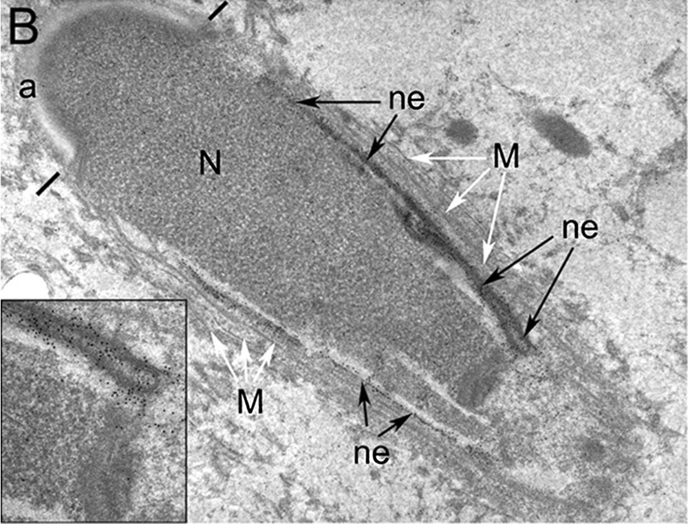
Human Elongated Spermatid Electron Micrograph
Elongated spermatid anti-SPANXa/d antibodies gold particles were associated with both the nucleoplasmic and the cytoplasmic faces of the nuclear envelope (ne) posterior to the acrosome (a) and adjacent to the manchette microtubules (M, white arrows). Staining of the nuclear envelope was not observed underlying the developing acrosome (a, bars mark posterior aspect of acrosome) or in the region immediately posterior to the acrosomal bulbs where the nuclear envelope is closely apposed to the nucleus.
The inset shows a fold of the nuclear envelope at the posterior region of the spermatid nucleus which exhibited a concentration of gold particles indicative of SPANXa/d staining. M, manchette; N, nucleus.
B × 16,000.
- Human Spermatozoa EM: Image - cap-phase spermatid | Image - elongated spermatid | Image - spermatid | Image - spermatozoa | Image - normal nucleus | Image - nucleus | Image - abnormal nucleus | Spermatozoa Development | Category:Electron Micrograph
- Spermatozoa Images: Spermatozoa BF | Spermatozoon BF | Spermatozoon EM | Spermatozoon EM | Historic drawing | Category:Spermatozoa | Spermatozoa Development | Testis Development
Reference
Westbrook VA, Schoppee PD, Vanage GR, Klotz KL, Diekman AB, Flickinger CJ, Coppola MA & Herr JC. (2006). Hominoid-specific SPANXA/D genes demonstrate differential expression in individuals and protein localization to a distinct nuclear envelope domain during spermatid morphogenesis. Mol. Hum. Reprod. , 12, 703-16. PMID: 17012309 DOI.
Copyright
© The Author 2006. Published by Oxford University Press on behalf of the European Society of Human Reproduction and Embryology. All rights reserved. For Permissions, please email: journals.permissions@oxfordjournals.org
The online version of this article has been published under an open access model. Users are entitled to use, reproduce, disseminate, or display the open access version of this article for non-commercial purposes provided that: the original authorship is properly and fully attributed; the Journal and Oxford University Press are attributed as the original place of publication with the correct citation details given; if an article is subsequently reproduced or disseminated not in its entirety but only in part or as a derivative work this must be clearly indicated. For commercial re-use, please contact journals.permissions@oxfordjournals.org
Original file name: Figure 8. http://molehr.oxfordjournals.org/content/12/11/703/F8.expansion.html Panel B cropped and resized from original figure.
Cite this page: Hill, M.A. (2024, April 19) Embryology Human spermatid EM02.jpg. Retrieved from https://embryology.med.unsw.edu.au/embryology/index.php/File:Human_spermatid_EM02.jpg
- © Dr Mark Hill 2024, UNSW Embryology ISBN: 978 0 7334 2609 4 - UNSW CRICOS Provider Code No. 00098G
File history
Click on a date/time to view the file as it appeared at that time.
| Date/Time | Thumbnail | Dimensions | User | Comment | |
|---|---|---|---|---|---|
| current | 10:15, 17 September 2014 | | 1,000 × 762 (186 KB) | Z8600021 (talk | contribs) | ==Human Spermatid Electron Micrograph== Electron micrographs of human spermatids following post-embedding labelling with anti-SPANXa/d antibodies. Elongated spermatid gold particles were associated with both the nucleoplasmic and the cytoplasmic fac... |
You cannot overwrite this file.
File usage
The following page uses this file: