File:Human ovary non-growing follicle model.jpg

Original file (1,151 × 679 pixels, file size: 116 KB, MIME type: image/jpeg)
Human Ovary Non-Growing Follicle Model
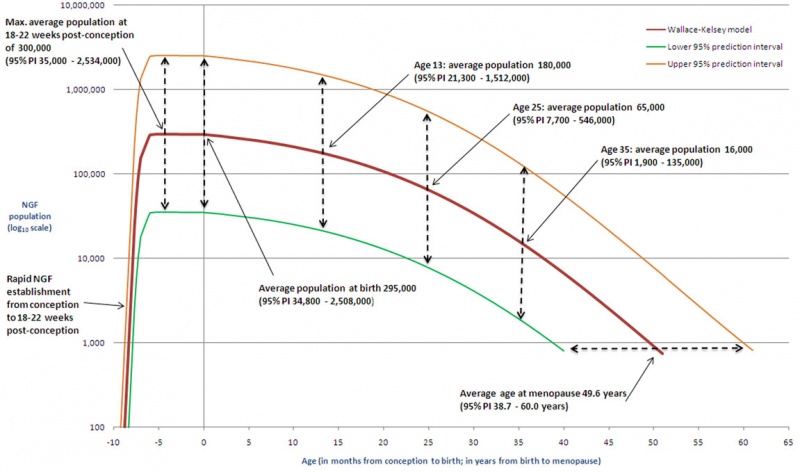
This figure gives illustrative examples of non-growing follicle (NGF, ovarian reserve) populations predicted by the original paper's model.[1]
- At ages 20 weeks, birth, 13 years, 25 years and 35 years the average NGF population is given, together with the respective 95% prediction intervals.
- The predicted average age at menopause (49.6 years) is also shown, together with the 95% prediction interval.
- The human ovary contains a fixed number of non-growing follicles (NGFs) established before birth that decline with increasing age culminating in the menopause at 50–51 years.
See also earlier oocyte number calculation.
- Links: ovary | oocyte | Human ovary non-growing follicle model 02.jpg | File:Human ovary non-growing follicle model.jpg
Ovarian Reserve
The ovarian reserve dogma in human (mammalian) development is that new oocyte and follicle production does not occur during postnatal life. A recently published human ovary molecular study has reinforced this dogma.[2] There is also substantial data that shows human ovarian changes postnatally are loss by apoptosis of prenatal oocytes.
Reference
- ↑ Wallace WH & Kelsey TW. (2010). Human ovarian reserve from conception to the menopause. PLoS ONE , 5, e8772. PMID: 20111701 DOI.
- ↑ Wagner M, Yoshihara M, Douagi I, Damdimopoulos A, Panula S, Petropoulos S, Lu H, Pettersson K, Palm K, Katayama S, Hovatta O, Kere J, Lanner F & Damdimopoulou P. (2020). Single-cell analysis of human ovarian cortex identifies distinct cell populations but no oogonial stem cells. Nat Commun , 11, 1147. PMID: 32123174 DOI.
Copyright
© 2010 Wallace, Kelsey. This is an open-access article distributed under the terms of the Creative Commons Attribution License, which permits unrestricted use, distribution, and reproduction in any medium, provided the original author and source are credited.
Figure 4. Illustrative examples. Journal.pone.0008772.g004.jpg
Cite this page: Hill, M.A. (2024, April 26) Embryology Human ovary non-growing follicle model.jpg. Retrieved from https://embryology.med.unsw.edu.au/embryology/index.php/File:Human_ovary_non-growing_follicle_model.jpg
- © Dr Mark Hill 2024, UNSW Embryology ISBN: 978 0 7334 2609 4 - UNSW CRICOS Provider Code No. 00098G
File history
Click on a date/time to view the file as it appeared at that time.
| Date/Time | Thumbnail | Dimensions | User | Comment | |
|---|---|---|---|---|---|
| current | 16:19, 17 May 2011 | | 1,151 × 679 (116 KB) | S8600021 (talk | contribs) | ==Human ovary non-growing follicle model== This figure gives illustrative examples of non-growing follicle (NGF) populations predicted by our model. At ages 20 weeks, birth, 13 years, 25 years and 35 years the average NGF population is given, together wi |
You cannot overwrite this file.
File usage
The following 12 pages use this file:
- ANAT2341 Lab 1 - Oogenesis
- BGDA Practical 3 - Oogenesis and Ovulation
- BGD Lecture - Sexual Differentiation
- Fertilization
- In Vitro Oogenesis
- Lecture - Genital Development
- Menstrual Cycle
- Oocyte Development
- Ovary Development
- REI - Reproductive Medicine Seminar 2018
- Royal Hospital for Women - Reproductive Medicine Seminar 2018
- Timeline human development