File:Human-spermatozoa EM01.jpg
Original file (1,000 × 204 pixels, file size: 26 KB, MIME type: image/jpeg)
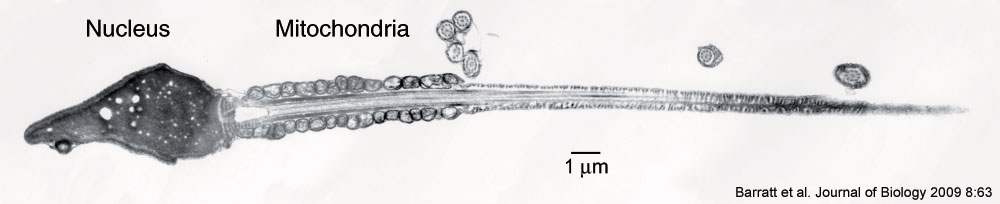
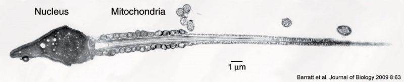
Human Spermatozoa ( transmission electron micrograph)
Montage transmission electron micrograph of a human sperm cell.
The cell has a compact nucleus, conspicuous mitochondria, no endoplasmic reticulum, minimal cytoplasm and a large tail (about 45 μm in length). Superfluous cytoplasm and associated machinery is jettisoned when the sperm emerges from the testis, leaving a 'stripped down', minimalist cell.
- Cell membrane - containing membrane proteins for chemotaxis and binding to the oocyte zone pellucida.
- Acrosome - containing enzymes required to digest the zona pellucida. The acrosome develops as a highly modified golgi structure.
- Nucleus - containing male haploid genome required to combine with oocyte haploid genome to form diploid zygote.
- Neck - Centriole and axonema required for spermatozoa movement.
- Middle piece - Mitochondria and annulus required for energy for spermatozoa movement.
- Principal piece - Microtubules required for spermatozoa movement.
- Human Spermatozoa EM: Image - cap-phase spermatid | Image - elongated spermatid | Image - spermatid | Image - spermatozoa | Image - normal nucleus | Image - nucleus | Image - abnormal nucleus | Spermatozoa Development | Category:Electron Micrograph
- Spermatozoa Images: Spermatozoa BF | Spermatozoon BF | Spermatozoon EM | Spermatozoon EM | Historic drawing | Category:Spermatozoa | Spermatozoa Development | Testis Development
Reference
<pubmed>19678911</pubmed>| PMC: 2736672 | J of Biology
BioMed Central Open Access BioMed Central Open Access license agreement Brief summary of the agreement.
Anyone is free: to copy, distribute, and display the work; to make derivative works; to make commercial use of the work.
Under the following conditions: Attribution, the original author must be given credit; for any reuse or distribution, it must be made clear to others what the license terms of this work are; any of these conditions can be waived if the authors gives permission.
Barratt et al. Journal of Biology 2009 8:63 doi:10.1186/jbiol167
File history
Click on a date/time to view the file as it appeared at that time.
| Date/Time | Thumbnail | Dimensions | User | Comment | |
|---|---|---|---|---|---|
| current | 13:24, 5 April 2010 | 1,000 × 204 (26 KB) | S8600021 (talk | contribs) | Montage transmission electron micrograph of a human sperm cell. The cell has a compact nucleus, conspicuous mitochondria, no endoplasmic reticulum, minimal cytoplasm and a large tail (about 45 μm in length). Superfluous cytoplasm and associated machine |
You cannot overwrite this file.