File:Horseshoe kidney.jpg
From Embryology
No higher resolution available.
Horseshoe_kidney.jpg (776 × 416 pixels, file size: 39 KB, MIME type: image/jpeg)
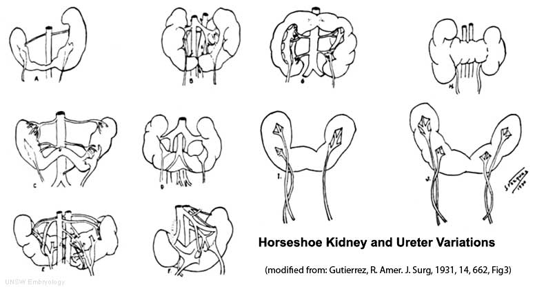
Renal Fusion Abnormality - Horseshoe Kidney
In the horseshoe kidney there is typically a fusion of the lower poles of both kidneys.
- During migration from the sacral region the two metanephric blastemas can come into contact mainly at the lower pole.
- The ureters pass in front of the zone of fusion of the kidneys.
- The kidneys and ureters usually function reasonably.
- Increased incidence of upper urinary tract obstruction or infection.
- Some horseshoe variations have been described as having associated ureter abnormalities including duplications.
Cite this page: Hill, M.A. (2024, April 19) Embryology Horseshoe kidney.jpg. Retrieved from https://embryology.med.unsw.edu.au/embryology/index.php/File:Horseshoe_kidney.jpg
- © Dr Mark Hill 2024, UNSW Embryology ISBN: 978 0 7334 2609 4 - UNSW CRICOS Provider Code No. 00098G
File history
Click on a date/time to view the file as it appeared at that time.
| Date/Time | Thumbnail | Dimensions | User | Comment | |
|---|---|---|---|---|---|
| current | 13:55, 19 September 2009 | | 776 × 416 (39 KB) | S8600021 (talk | contribs) |
You cannot overwrite this file.