File:Gray1202.jpg
Gray1202.jpg (563 × 500 pixels, file size: 63 KB, MIME type: image/jpeg)
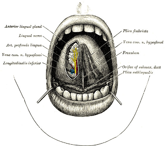
The Oral (Mouth) Cavity
The apex of the tongue is turned upward, and on the right side a superficial dissection of its under surface has been made.
On the under surface of the tongue the mucous membrane is smooth and devoid of papillæ. In the middle line, the mucous membrane extends to the floor of the mouth as a distinct fold—the frenulum—the free edge of which runs forward to the symphysis menti. Sometimes the ranine vein can be seen immediately beneath the mucous membrane, a little lateral to the frenulum.
Close to the attachment of the frenulum to the floor of the mouth, the slit-like orifice of the submaxillary duct is visible on either side. Running backward and lateralward from the orifice of the submaxillary duct is the plica sublingualis, produced by the projection of the sublingual gland which lies immediately beneath the mucous membrane. The plica serves also to indicate the line of the submaxillary duct and of the lingual nerve.
At the back of the mouth is the isthmus faucium, bounded above by the palatine velum, from the free margin of which the uvula projects downward in the middle line.
On either side of the isthmus are the two palatine arches, the anterior formed by the Glossopalatinus and the posterior by the Pharyngopalatinus. Between the two arches of either side is the palatine tonsil, above which is the small supratonsillar recess; the position of the tonsil corresponds to the angle of the mandible.
When the mouth is opened widely, a tense band—the pterygomandibular raphé—can be seen and felt lateral to the glossopalatine arch. Its lower end is attached to the mandible behind the last molar tooth, and immediately below and in front of this the lingual nerve can be felt; the upper end of the ligament can be traced to the pterygoid hamulus. About 1 cm. in front of the hamulus and 1 cm. medial to the last molar tooth of the maxilla is the greater palatine foramen through which the descending palatine vessels and the anterior palatine nerve emerge. Behind the last molar tooth of the maxilla the coronoid process of the mandible is palpable.
- Gray's Images: Development | Lymphatic | Neural | Vision | Hearing | Somatosensory | Integumentary | Respiratory | Gastrointestinal | Urogenital | Endocrine | Surface Anatomy | iBook | Historic Disclaimer
| Historic Disclaimer - information about historic embryology pages |
|---|
 |
| iBook - Gray's Embryology | |
|---|---|

|
|
Reference
Gray H. Anatomy of the human body. (1918) Philadelphia: Lea & Febiger.
Cite this page: Hill, M.A. (2024, April 26) Embryology Gray1202.jpg. Retrieved from https://embryology.med.unsw.edu.au/embryology/index.php/File:Gray1202.jpg
- © Dr Mark Hill 2024, UNSW Embryology ISBN: 978 0 7334 2609 4 - UNSW CRICOS Provider Code No. 00098G
File history
Click on a date/time to view the file as it appeared at that time.
| Date/Time | Thumbnail | Dimensions | User | Comment | |
|---|---|---|---|---|---|
| current | 06:23, 23 November 2010 | | 563 × 500 (63 KB) | S8600021 (talk | contribs) | ==The Oral (Mouth) Cavity== The apex of the tongue is turned upward, and on the right side a superficial dissection of its under surface has been made. On the under surface of the tongue the mucous membrane is smooth and devoid of papillæ. In the midd |
You cannot overwrite this file.
File usage
The following 2 pages use this file: