File:Gray1161.jpg
Original file (1,000 × 671 pixels, file size: 138 KB, MIME type: image/jpeg)
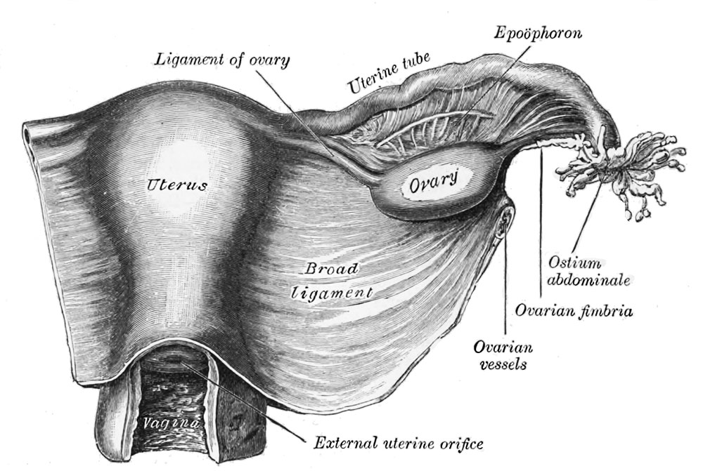
Uterus and Right Broad Ligament
Uterus and right broad ligament, seen from behind. The broad ligament has been spread out and the ovary drawn downward.
The ovaries are homologous with the testes in the male. They are two nodular bodies, situated one on either side of the uterus in relation to the lateral wall of the pelvis, and attached to the back of the broad ligament of the uterus, behind and below the uterine tubes (Fig. 1161).
The ovaries are of a grayish-pink color, and present either a smooth or a puckered uneven surface. They are each about 4 cm. in length, 2 cm. in width, and about 8 mm. in thickness, and weigh from 2 to 3.5 gm. Each ovary presents a lateral and a medial surface, an upper or tubal and a lower or uterine extremity, and an anterior or mesovarion and a posterior free border. It lies in a shallow depression, named the ovarian fossa, on the lateral wall of the pelvis; this fossa is bounded above by the external iliac vessels, in front by the obliterated umbilical artery, and behind by the ureter. The exact position of the ovary has been the subject of considerable difference of opinion, and the description here given applies to the ovary of the nulliparous woman. The ovary becomes displaced during the first pregnancy, and probably never again returns to its original position. In the erect posture the long axis of the ovary is vertical.
The tubal extremity is near the external iliac vein; to it are attached the ovarian fimbria of the uterine tube and a fold of peritoneum, the suspensory ligament of the ovary, which is directed upward over the iliac vessels and contains the ovarian vessels. The uterine end is directed downward toward the pelvic floor, it is usually narrower than the tubal, and is attached to the lateral angle of the uterus, immediately behind the uterine tube, by a rounded cord termed the ligament of the ovary, which lies within the broad ligament and contains some non-striped, muscular fibers. The lateral surface is in contact with the parietal peritoneum, which lines the ovarian fossa; the medial surface is to a large extent covered by the fimbriated extremity of the uterine tube. The mesovarian border is straight and is directed toward the obliterated umbilical artery, and is attached to the back of the broad ligament by a short fold named the mesovarium. Between the two layers of this fold the blood vessels and nerves pass to reach the hilum of the ovary. The free border is convex, and is directed toward the ureter. The uterine tube arches over the ovary, running upward in relation to its mesovarian border, then curving over its tubal pole, and finally passing downward on its free border and medial surface.
- Links: Uterus Development | Ovary Development
- Gray's Images: Development | Lymphatic | Neural | Vision | Hearing | Somatosensory | Integumentary | Respiratory | Gastrointestinal | Urogenital | Endocrine | Surface Anatomy | iBook | Historic Disclaimer
| Historic Disclaimer - information about historic embryology pages |
|---|
 |
| iBook - Gray's Embryology | |
|---|---|

|
|
Reference
Gray H. Anatomy of the human body. (1918) Philadelphia: Lea & Febiger.
Cite this page: Hill, M.A. (2024, April 24) Embryology Gray1161.jpg. Retrieved from https://embryology.med.unsw.edu.au/embryology/index.php/File:Gray1161.jpg
- © Dr Mark Hill 2024, UNSW Embryology ISBN: 978 0 7334 2609 4 - UNSW CRICOS Provider Code No. 00098G
File history
Click on a date/time to view the file as it appeared at that time.
| Date/Time | Thumbnail | Dimensions | User | Comment | |
|---|---|---|---|---|---|
| current | 10:18, 11 February 2014 | | 1,000 × 671 (138 KB) | Z8600021 (talk | contribs) | |
| 09:49, 11 February 2014 |  | 800 × 531 (104 KB) | Z8600021 (talk | contribs) | ==Uterus and Right Broad Ligament== Uterus and right broad ligament, seen from behind. The broad ligament has been spread out and the ovary drawn downward. {{Gray Anatomy}} Category:Uterus |
You cannot overwrite this file.
File usage
The following 4 pages use this file: