File:Gray1140.jpg
From Embryology
Size of this preview: 546 × 600 pixels. Other resolution: 600 × 659 pixels.
Original file (600 × 659 pixels, file size: 100 KB, MIME type: image/jpeg)
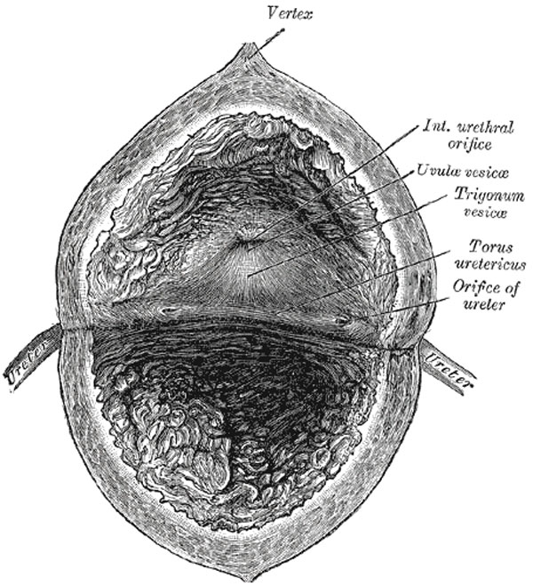
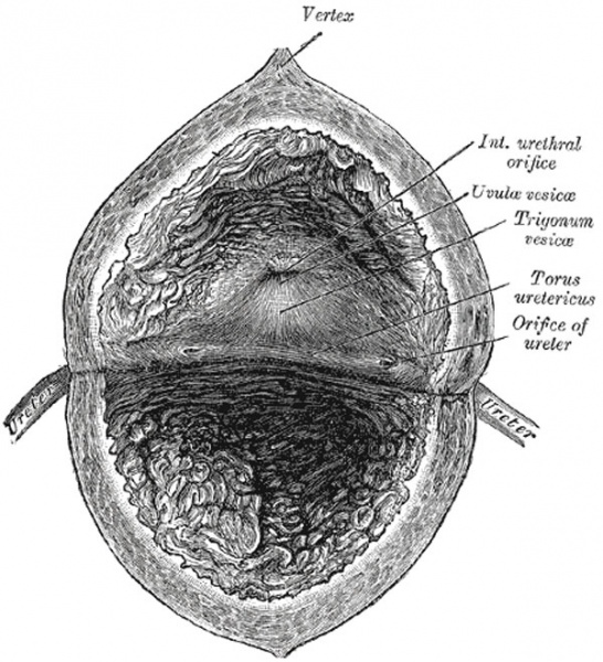
Fig. 1140. Interior of the Bladder
- The mucous membrane lining the bladder is, over the greater part of the viscus, loosely attached to the muscular coat, and appears wrinkled or folded when the bladder is contracted: in the distended condition of the bladder the folds are effaced.
- Over a small triangular area, termed the trigonum vesicæ, immediately above and behind the internal orifice of the urethra, the mucous membrane is firmly bound to the muscular coat, and is always smooth.
- The anterior angle of the trigonum vesicæ is formed by the internal orifice of the urethra: its postero-lateral angles by the orifices of the ureters. Stretching behind the latter openings is a slightly curved ridge, the torus uretericus, forming the base of the trigone and produced by an underlying bundle of non-striped muscular fibers.
- The lateral parts of this ridge extend beyond the openings of the ureters, and are named the plicæ uretericæ; they are produced by the terminal portions of the ureters as they traverse obliquely the bladder wall.
- When the bladder is illuminated the torus uretericus appears as a pale band and forms an important guide during the operation of introducing a catheter into the ureter.
- The orifices of the ureters are placed at the postero-lateral angles of the trigonum vesicæ, and are usually slit-like in form.
- In the contracted bladder they are about 2.5 cm. apart and about the same distance from the internal urethral orifice; in the distended viscus these measurements may be increased to about 5 cm.
- The internal urethral orifice is placed at the apex of the trigonum vesicæ, in the most dependent part of the bladder, and is usually somewhat crescentic in form; the mucous membrane immediately behind it presents a slight elevation, the uvula vesicæ, caused by the middle lobe of the prostate.
- Gray's Images: Development | Lymphatic | Neural | Vision | Hearing | Somatosensory | Integumentary | Respiratory | Gastrointestinal | Urogenital | Endocrine | Surface Anatomy | iBook | Historic Disclaimer
| Historic Disclaimer - information about historic embryology pages |
|---|
 |
| iBook - Gray's Embryology | |
|---|---|

|
|
Reference
Gray H. Anatomy of the human body. (1918) Philadelphia: Lea & Febiger.
Cite this page: Hill, M.A. (2024, April 18) Embryology Gray1140.jpg. Retrieved from https://embryology.med.unsw.edu.au/embryology/index.php/File:Gray1140.jpg
- © Dr Mark Hill 2024, UNSW Embryology ISBN: 978 0 7334 2609 4 - UNSW CRICOS Provider Code No. 00098G
File history
Click on a date/time to view the file as it appeared at that time.
| Date/Time | Thumbnail | Dimensions | User | Comment | |
|---|---|---|---|---|---|
| current | 22:23, 28 October 2010 | | 600 × 659 (100 KB) | S8600021 (talk | contribs) |
You cannot overwrite this file.
File usage
The following 3 pages use this file: