File:Gray0026.jpg
From Embryology

No higher resolution available.
Gray0026.jpg (500 × 500 pixels, file size: 37 KB, MIME type: image/jpeg)
Fig. 26. Diagram showing stage of allantoic development with commencing constriction of the yolk-sac
Diagram showing later stage of allantoic development with commencing constriction of the yolk-sac.
--Mark Hill (talk) 12:38, 25 April 2013 (EST)
- yolk sac - An extraembryonic membrane which is endoderm origin and covered with extraembryonic mesoderm. Yolk sac lies outside the embryo connected initially by a yolk stalk to the midgut with which it is continuous with. The endodermal lining is continuous with the endoderm of the gastrointestinal tract. The extra-embryonic mesoderm differentiates to form both blood and blood vessels of the vitelline system. In reptiles and birds, the yolk sac has a function associated with nutrition. In mammals the yolk sac acts as a source of primordial germ cells and blood cells. Note that in early development (week 2) a structure called the "primitive yolk sac" forms from hypoblast, this is an entirely different structure.
Early embryo membrane development cartoons: Image 24 | Image 25 | Image 26 | Image 27 | Image 28
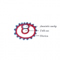
Fig 24
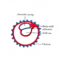
Fig 25
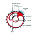
Fig 26
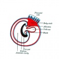
Fig 27
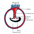
Fig 28


- Gray's Images: Development | Lymphatic | Neural | Vision | Hearing | Somatosensory | Integumentary | Respiratory | Gastrointestinal | Urogenital | Endocrine | Surface Anatomy | iBook | Historic Disclaimer
| Historic Disclaimer - information about historic embryology pages |
|---|
 |
| iBook - Gray's Embryology | |
|---|---|

|
|
Reference
Gray H. Anatomy of the human body. (1918) Philadelphia: Lea & Febiger.
Cite this page: Hill, M.A. (2024, April 19) Embryology Gray0026.jpg. Retrieved from https://embryology.med.unsw.edu.au/embryology/index.php/File:Gray0026.jpg
- © Dr Mark Hill 2024, UNSW Embryology ISBN: 978 0 7334 2609 4 - UNSW CRICOS Provider Code No. 00098G
File history
Click on a date/time to view the file as it appeared at that time.
| Date/Time | Thumbnail | Dimensions | User | Comment | |
|---|---|---|---|---|---|
| current | 11:53, 12 May 2013 | | 500 × 500 (37 KB) | Z8600021 (talk | contribs) |
You cannot overwrite this file.
File usage
The following 15 pages use this file:
- 2009 Lecture 8
- 2010 Lecture 8
- ANAT2341 Lab 4 - Implantation and Villi Development
- ASA Meeting 2013 - Placenta
- Anatomy of the Human Body by Henry Gray
- BGDA Practical Placenta - Implantation and Early Placentation
- Coelomic Cavity Development
- Lecture - Placenta Development
- File:Gray0024-29.gif
- File:Gray0024.jpg
- File:Gray0025.jpg
- File:Gray0026.jpg
- File:Gray0027.jpg
- File:Gray0028.jpg
- Template:Gray fetal membrane cartoons