File:Epidermis cartoon 02.jpg
Epidermis_cartoon_02.jpg (452 × 536 pixels, file size: 78 KB, MIME type: image/jpeg)
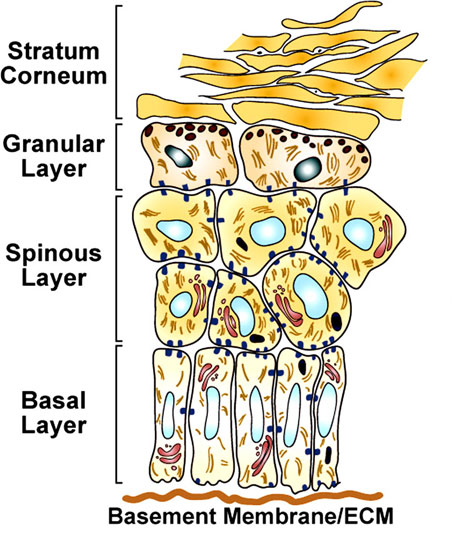
Epidermal Differentiation
The program of epidermal differentiation is shown in this schematic, illustrating the basement membrane at the base, the proliferative basal layer, and the three differentiation stages: spinous layer, granular layer, and outermost stratum corneum.
Related Image: same image with layer molecular information
Reference
Fuchs E. (2008). Skin stem cells: rising to the surface. J. Cell Biol. , 180, 273-84. PMID: 18209104 DOI.
Copyright
Rockefeller University Press - Copyright Policy This article is distributed under the terms of an Attribution–Noncommercial–Share Alike–No Mirror Sites license for the first six months after the publication date (see http://www.jcb.org/misc/terms.shtml). After six months it is available under a Creative Commons License (Attribution–Noncommercial–Share Alike 4.0 Unported license, as described at https://creativecommons.org/licenses/by-nc-sa/4.0/ ). (More? Help:Copyright Tutorial)
Cite this page: Hill, M.A. (2024, April 19) Embryology Epidermis cartoon 02.jpg. Retrieved from https://embryology.med.unsw.edu.au/embryology/index.php/File:Epidermis_cartoon_02.jpg
- © Dr Mark Hill 2024, UNSW Embryology ISBN: 978 0 7334 2609 4 - UNSW CRICOS Provider Code No. 00098G
Original file name: Figure 1. http://jcb.rupress.org/content/180/2/273/F1.expansion.html (resized and molecular information cropped)
File history
Click on a date/time to view the file as it appeared at that time.
| Date/Time | Thumbnail | Dimensions | User | Comment | |
|---|---|---|---|---|---|
| current | 12:36, 13 October 2010 | | 452 × 536 (78 KB) | S8600021 (talk | contribs) | ==Epidermal Differentiation== The program of epidermal differentiation is shown in this schematic, illustrating the basement membrane at the base, the proliferative basal layer, and the three differentiation stages: spinous layer, granular layer, and out |
You cannot overwrite this file.
File usage
The following 2 pages use this file: