File:Endochondral ossification.jpg
Endochondral_ossification.jpg (400 × 533 pixels, file size: 91 KB, MIME type: image/jpeg)
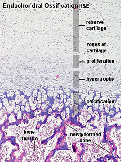
Endochondral Ossification
This process of developmental bone formation is the replacement of an earlier cartilage "template" with bone.
In this histological image, the cartilage is shown at the top of the image and the newly formed bone is at the bottom of the image.
- Bone Histology: Cartilage Histology | Histology Stains | Histology | cartilage | bone | bone timeline
- Trabecular bone trabecular | lamellar | trabecular - overview HE | trabecular - low HE | trabecular - med HE
- Endochondral ossification primary ossification | endochondral ossification
- Intramembranous ossification intramembranous - VG low | intramembranous - VG high | intramembranous - HE low | intramembranous - HE high
Links: Histology | Histology Stains | Blue Histology images copyright Lutz Slomianka 1998-2009. The literary and artistic works on the original Blue Histology website may be reproduced, adapted, published and distributed for non-commercial purposes. See also the page Histology Stains.
Cite this page: Hill, M.A. (2024, April 19) Embryology Endochondral ossification.jpg. Retrieved from https://embryology.med.unsw.edu.au/embryology/index.php/File:Endochondral_ossification.jpg
- © Dr Mark Hill 2024, UNSW Embryology ISBN: 978 0 7334 2609 4 - UNSW CRICOS Provider Code No. 00098G
File history
Click on a date/time to view the file as it appeared at that time.
| Date/Time | Thumbnail | Dimensions | User | Comment | |
|---|---|---|---|---|---|
| current | 20:12, 15 March 2015 | | 400 × 533 (91 KB) | Z8600021 (talk | contribs) | |
| 11:33, 11 September 2009 |  | 300 × 400 (73 KB) | S8600021 (talk | contribs) | Enos04he.jpg |
You cannot overwrite this file.
File usage
The following 15 pages use this file:
- 2009 Lecture 13
- 2010 BGD Lecture - Development of the Embryo/Fetus 2
- 2010 BGD Practical 6 - Week 7
- 2010 Lecture 13
- ANAT2241 Bone, Bone Formation and Joints
- ANAT2241 Connective Tissue Components
- ANAT2241 Connective Tissue Types
- BGDA Lecture - Development of the Embryo/Fetus 2
- BGDA Practical 7 - Week 7
- Bone Development
- Bone Histology
- Cartilage Histology
- E
- Lecture - Musculoskeletal Development
- Musculoskeletal System - Bone Development