File:Blood capillary EM 02.jpg
Blood_capillary_EM_02.jpg (600 × 600 pixels, file size: 99 KB, MIME type: image/jpeg)
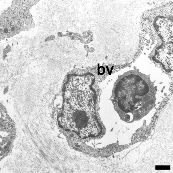
Blood Capillary Electron Micrograph
A blood capillary with white blood cell (monocyte) located in the lumen.
Note the nuclei of the endothelial cells bulging into the vessel lumen.
The white blood cell nucleus shape suggests that it is a Monocyte.
Scale bar 1 μm (Stain - Osmium)
- Capillary EM Histology Links: Capillary 1 large unlabeled | Capillary 1 large labeled | Virtual Slide | Capillary 1 small unlabeled | Capillary 1 small labeled | endothelium detail | Containing white blood cell | Blood Vessel Histology | Medicine - Histology | Blood Vessel Development
- Blood Histology: Blood Development | Blood Cell Number Table | Lymphocyte 1 | Lymphocyte 2 | Lymphocyte 3 | Lymphocyte 4 | Monocyte 1 | Monocyte 2 | Monocyte 3 | Monocyte 4 | Neutrophils 1 | Neutrophil 2 | Neutrophil 3 | Neutrophil 4 | Eosinophil 1 | Eosinophil 2 | labeled Neutrophil and Eosinophil | unlabeled - Neutrophil and Eosinophil | Basophil 1 | Basophil 2 | Basophil 3 | Platelet 1 | Platelet 2 | Reticulocyte | Megakaryocyte | Movie | Bone Marrow Histology | Category:Blood
| Blood Cells |
|---|
Adult human blood cell numbers shown in the table below is for reference purposes.
Blood Cell NumbersThe adult ranges of cells / 1 litre (l), total blood volume is about 4.7 to 5 litres. Blood Development | Blood Histology Red Blood Cells
Leukocytes (white blood cells)
Granulocytes
Non-Granulocytes
Lymphocytes
Platelets
|
Reference
Detry B, Bruyère F, Erpicum C, Paupert J, Lamaye F, Maillard C, Lenoir B, Foidart JM, Thiry M & Noël A. (2011). Digging deeper into lymphatic vessel formation in vitro and in vivo. BMC Cell Biol. , 12, 29. PMID: 21702933 DOI.
Detry et al. BMC Cell Biology 2011 12:29 doi:10.1186/1471-2121-12-29
Copyright
© 2011 Detry et al; licensee BioMed Central Ltd.
This is an Open Access article distributed under the terms of the Creative Commons Attribution License (http://creativecommons.org/licenses/by/2.0), which permits unrestricted use, distribution, and reproduction in any medium, provided the original work is properly cited.
Original file name: Panel C cropped from Figure 4. (1471-2121-12-29-4.jpg) Contrast and size adjusted.
Cite this page: Hill, M.A. (2024, April 16) Embryology Blood capillary EM 02.jpg. Retrieved from https://embryology.med.unsw.edu.au/embryology/index.php/File:Blood_capillary_EM_02.jpg
- © Dr Mark Hill 2024, UNSW Embryology ISBN: 978 0 7334 2609 4 - UNSW CRICOS Provider Code No. 00098G
File history
Click on a date/time to view the file as it appeared at that time.
| Date/Time | Thumbnail | Dimensions | User | Comment | |
|---|---|---|---|---|---|
| current | 10:43, 5 February 2012 | | 600 × 600 (99 KB) | S8600021 (talk | contribs) | ==Blood Capillary Electron Micrograph== A blood capillary with white blood cell in lumen. Scale bar 1 μm Panel C cropped from Figure 4. (1471-2121-12-29-4.jpg) Contrast and size adjusted. ===Reference=== <pubmed>21702933</pubmed>| [http://www.ncbi.nl |
You cannot overwrite this file.
File usage
The following 3 pages use this file: