File:Bailey355.jpg
Original file (632 × 890 pixels, file size: 109 KB, MIME type: image/jpeg)
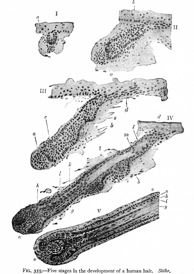
Fig. 355. Five stages in the development of a human hair
Stohr.
The hairs are derivatives of the epidermal layer of the ectoderm.
- I-II - about three months, local thickenings of the epidermis appear (beginning in the region of the forehead and eye-brows) and grow obliquely into the underlying dermis in the form of solid buds the hair germs. As the buds continue to elongate they become club-shaped and the epithelium at the end of each molds itself over a little portion of the dermis in which the cells have become more numerous and which is known as the hair papilla (Fig. 354).
- III - IV - As the epidermal bud grows deeper, its central cells become spindle-shaped and undergo keratinization to form the beginning of the hair shaft; the peripheral layers constitute the anlage of the root sheath. The hair shaft grows from its basal end, new keratinized cells being added from the epithelium nearest the papilla as the older cells are pushed toward the surface of the skin.
- V - The surface cells of the hair shaft become flattened to form the cuticle of the hair. The hairs appear above the surface about the fifth month. Of the cells of the root sheath, those nearest the hair become scale-like to form the cuticle of the root sheath; the next few layers become modified (keratinized) to form Huxley's and Henle's layers. Outside of these is the stratum germinativum, the basal layer of which is composed of columnar cells resting upon a distinct basement membrane. The stratum germinativum is continued over the tip of the papilla, where its cells give rise to new cells for the hair shaft.
The connective tissue around the root sheath becomes differentiated into an inner highly vascular layer, the fibers of which run circularly, and an outer layer, the fibers of which extend along the sheath. The two layers together constitute the connective tissue follicle.
The first formed hairs, which are exceedingly fine and silky, develop in vast numbers over the surface of the embryonic body and are known collectively as the lanugo. This growth is lost (beginning before birth and continuing during the first and second years after), except over the face, and is replaced by coarser hairs. These in turn are constantly being shed during the life of the individual and replaced by new ones. The new hairs probably in most cases develop from the old follicles, the cells over the old papillae proliferating and the newly formed hairs growing up through the old sheaths. In some cases, however, new follicles are formed directly from the epidermis and dermis. In some of the lower Mammals, new hair germs appear as outgrows from the sheaths of old follicles, thus giving rise to tufts of hair. The arrectores pilorum muscles arise from the dermal (mesenchymal) cells and become attached to the follicles below the sebaceous glands.
Legend
a, Papilla; b, arrector pili muscle; c, beginning of hair shaft; d, point where hair shaft grows through epidermis; e, anlage of sebaceous gland; /, hair germ; g, hair shaft; h, Henle's layer; i, Huxley's layer; k, cuticle of root sheath; l, inner root sheath; m, outer root sheath in tangential section; n, outer root sheath; o, connective tissue follicle.
- Text-Book of Embryology: Germ cells | Maturation | Fertilization | Amphioxus | Frog | Chick | Mammalian | External body form | Connective tissues and skeletal | Vascular | Muscular | Alimentary tube and organs | Respiratory | Coelom, Diaphragm and Mesenteries | Urogenital | Integumentary | Nervous System | Special Sense | Foetal Membranes | Teratogenesis | Gallery of All Figures
| Historic Disclaimer - information about historic embryology pages |
|---|
 |
Reference
Bailey FR. and Miller AM. Text-Book of Embryology (1921) New York: William Wood and Co.
Cite this page: Hill, M.A. (2024, April 18) Embryology Bailey355.jpg. Retrieved from https://embryology.med.unsw.edu.au/embryology/index.php/File:Bailey355.jpg
- © Dr Mark Hill 2024, UNSW Embryology ISBN: 978 0 7334 2609 4 - UNSW CRICOS Provider Code No. 00098G
File history
Click on a date/time to view the file as it appeared at that time.
| Date/Time | Thumbnail | Dimensions | User | Comment | |
|---|---|---|---|---|---|
| current | 15:59, 29 January 2011 | | 632 × 890 (109 KB) | S8600021 (talk | contribs) | ==Fig. 355. Five stages in the development of a human hair== Stohr. a, Papilla; b, arrector pili muscle; c, beginning of hair shaft; d, point where hair shaft grows through epidermis; e, anlage of sebaceous gland; /, hair germ; g, hair shaft; h, Henle' |
You cannot overwrite this file.
File usage
The following 2 pages use this file: