File:Bailey327.jpg
From Embryology

Size of this preview: 800 × 520 pixels. Other resolution: 872 × 567 pixels.
Original file (872 × 567 pixels, file size: 89 KB, MIME type: image/jpeg)
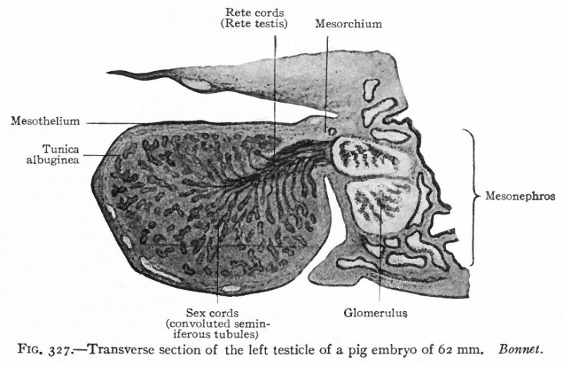
Fig. 327. Transverse section of the left testicle of a pig embryo of 62 mm
Bonnet.
After the fourth or fifth week, certain changes occur in the genital ridges which differ accordingly as the ridges form ovaries or testicles. While the differences are at first not particularly obvious, there are four which become clearer as the changes progress,
- If the ridge is to become a testicle, the cells of the surface epithelium become arranged in a single layer and become flat.
- In a developing testicle a layer of dense connective tissue grows between the surface epithelium and the sex cords, forming the tunica albuginea.
- In a testicle there also appears a sharper line of demarkation between the cell columns and the stroma, and the latter shows a more extensive growth.
- Another feature of the testicle is that the sex cells begin to be less conspicuous and do not increase further in size, but come to resemble the other epithelial elements.
- Text-Book of Embryology: Germ cells | Maturation | Fertilization | Amphioxus | Frog | Chick | Mammalian | External body form | Connective tissues and skeletal | Vascular | Muscular | Alimentary tube and organs | Respiratory | Coelom, Diaphragm and Mesenteries | Urogenital | Integumentary | Nervous System | Special Sense | Foetal Membranes | Teratogenesis | Gallery of All Figures
| Historic Disclaimer - information about historic embryology pages |
|---|
| Pages where the terms "Historic" (textbooks, papers, people, recommendations) appear on this site, and sections within pages where this disclaimer appears, indicate that the content and scientific understanding are specific to the time of publication. This means that while some scientific descriptions are still accurate, the terminology and interpretation of the developmental mechanisms reflect the understanding at the time of original publication and those of the preceding periods, these terms, interpretations and recommendations may not reflect our current scientific understanding. (More? Embryology History | Historic Embryology Papers) |
Reference
Bailey FR. and Miller AM. Text-Book of Embryology (1921) New York: William Wood and Co.
Cite this page: Hill, M.A. (2024, April 25) Embryology Bailey327.jpg. Retrieved from https://embryology.med.unsw.edu.au/embryology/index.php/File:Bailey327.jpg
- © Dr Mark Hill 2024, UNSW Embryology ISBN: 978 0 7334 2609 4 - UNSW CRICOS Provider Code No. 00098G
File history
Click on a date/time to view the file as it appeared at that time.
| Date/Time | Thumbnail | Dimensions | User | Comment | |
|---|---|---|---|---|---|
| current | 12:02, 25 January 2011 |  | 872 × 567 (89 KB) | S8600021 (talk | contribs) | {{Template:Bailey 1921 Figures}} Category:Human Category:Renal |
You cannot overwrite this file.
File usage
The following 5 pages use this file:
