File:3D Human pancreatic islet.jpg
From Embryology
Size of this preview: 510 × 600 pixels. Other resolution: 1,088 × 1,280 pixels.
Original file (1,088 × 1,280 pixels, file size: 295 KB, MIME type: image/jpeg)
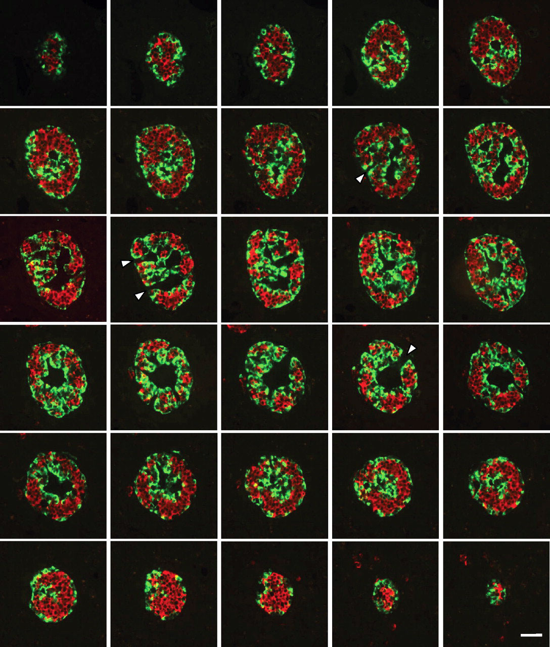
Three-dimensional Analysis of Human Pancreatic Islet
- Consecutive sections through an entire adult pancreatic islet labeled for insulin (red) and glucagon (green).
- Images show that insulin (red) and glucagon (green) stained cells are organized into continuous 3-D networks that span the entire islet.
- Sections at both ends show islet profiles with an apparent similar core-mantle structure to that of 40- to 60-μm–diameter islets.
- These consecutive images also reveal that vascular channels were in fact continuous ramified structures that were connected in places with the surrounding islet tissue (arrowheads).
- Image series is representative of 15 different pancreatic islets analyzed from one pancreas.
Scale bar, 50 μm.
Original file name: FIG. 4. http://www.ncbi.nlm.nih.gov/pmc/articles/PMC2857900/figure/F4/ http://diabetes.diabetesjournals.org/content/59/5/1202/F4.large.jpg
Reference
<pubmed>20185817</pubmed>| PMC2857900 | Diabetes.
© 2010 by the American Diabetes Association
Readers may use this article as long as the work is properly cited, the use is educational and not for profit, and the work is not altered. See http://creativecommons.org/licenses/by-nc-nd/3.0/ for details.
File history
Click on a date/time to view the file as it appeared at that time.
| Date/Time | Thumbnail | Dimensions | User | Comment | |
|---|---|---|---|---|---|
| current | 13:43, 8 August 2011 | | 1,088 × 1,280 (295 KB) | S8600021 (talk | contribs) | ==Three-dimensional Analysis of Human Pancreatic Islet== Consecutive sections through an entire pancreatic islet labeled for insulin (red) and glucagon (green). Images show that insulin (red)– and glucagon (green)–stained cells are organized into con |
You cannot overwrite this file.
File usage
The following 2 pages use this file: