Book - The Frog Its Reproduction and Development 12
| Embryology - 16 Apr 2024 |
|---|
| Google Translate - select your language from the list shown below (this will open a new external page) |
|
العربية | català | 中文 | 中國傳統的 | français | Deutsche | עִברִית | हिंदी | bahasa Indonesia | italiano | 日本語 | 한국어 | မြန်မာ | Pilipino | Polskie | português | ਪੰਜਾਬੀ ਦੇ | Română | русский | Español | Swahili | Svensk | ไทย | Türkçe | اردو | ייִדיש | Tiếng Việt These external translations are automated and may not be accurate. (More? About Translations) |
Rugh R. Book - The Frog Its Reproduction and Development. (1951) The Blakiston Company.
| Historic Disclaimer - information about historic embryology pages |
|---|
| Pages where the terms "Historic" (textbooks, papers, people, recommendations) appear on this site, and sections within pages where this disclaimer appears, indicate that the content and scientific understanding are specific to the time of publication. This means that while some scientific descriptions are still accurate, the terminology and interpretation of the developmental mechanisms reflect the understanding at the time of original publication and those of the preceding periods, these terms, interpretations and recommendations may not reflect our current scientific understanding. (More? Embryology History | Historic Embryology Papers) |
Chapter 12 - The Endodermal Derivatives
The endoderm gives rise to all structures associated with the original archenteron, from the mouth (ectodermal stomodeum) to the anus (ectodermal proctodeum), and all of its derivatives. It must be emphasized, however, that the endoderm contributes only the lining epithelium of these structures and that (since each of them is invested with blood, connective and nervous tissue, and often with muscle) ectoderm and mesoderm may also be involved. It is nevertheless convenient to describe in sequence from the anterior to the posterior (foregut, midgut, and hindgut) those structures whose linings are endodermal in origin.
The Mouth
(Stomodeum, Jaws, Lips, Etc.)
The lips and anterior lining of the mouth are ectodermal and the margin between the ectoderm and endoderm can be determined in the larva by the extent of the invaginated and pigmented stomodeal ectoderm. This ectoderm plus the oral endoderm form the oral plate, or oral membrane, which generally ruptures through shortly after the time of hatching (6 mm. stage) to form the mouth opening. The lateral margins of the mouth (stomodeum) are the original mandibular ridges between which are the dorsal and the ventral (larval) lips. These are transitory but important feeding organs of the tadpole. The dorsal lip develops three medially placed rows of superficial teeth which are periodically sloughed off and replaced. The ventral lip of the larva develops four rows of somewhat more complete teeth, but all teeth are covered with stomodeal ectoderm. A cornified ectodermal "jaw," consisting of a hardened ridge, develops at the base of both the dorsal and the ventral lips.
By the time of metamorphosis the horny larval teeth and jaws are lost and the mandibular arches give rise to the jaw elements of the adult. The upper larval teeth are replaced by permanent teeth which bear only superficial resemblance to the teeth of mammals.
The tongue, which is ultimately attached anteriorly and will be free posteriorly, begins to develop by the time of metamorphosis from a proliferation of cells from the endodermal floor of the pharynx. The bulk of the tongue will, of course, be of mesodermal origin but most of its covering and glandular elements come from the endoderm, the anterior portion being covered with stomodeal ectoderm.
The Foregut
This anterior portion of the original archenteron expands widely in front of the yolk mass and consists of all of the derivatives from the stomodeum to and including the pancreas and liver. The stomodeum, the anterior covering of the tongue, and the roof of the mouth anterior to the internal choanae are ectodermal and only the posterior covering of the tongue and mouth, beginning at about the level of the thyroid gland, is endodermal. This foregut portion of the archenteron therefore assumes a major role in the early development of the frog, where it gives rise to three successive sets of respiratory organs (i.e., external and internal gills, then the lungs) to most of the endocrine organs, and to a good portion of the digestive tract.
The Pharynx
The pharyngeal cavity is widely expanded anterior and ventral to the yolk mass of the frog embryo. The earliest elongated and vertical evaginations of the pharyngeal endoderm are known as the visceral or branchial pouches. Those of the most anterior pair, which develop between the mandibular and the hyoid arches, are known as the hyomandibular pouches. The dorsal remnant of this first pair of pouches gives rise to the Eustachian tube of the adult and connects the pharynx with the middle ear chamber. The second and third pairs of visceral pouches are known as the first and second branchial pouches, because they give rise to gill clefts, and they develop between the succeeding visceral arches. Eventually, six pairs of endodermal evaginations develop in this manner, but those of the last (most posterior) pair rarely open as pouches, remaining as mere cords of endoderm. By definition we use the term "pouch" for the endodermal evagination, "groove" for the ectodermal invagination, and "cleft" for the combination of ectoderm and endoderm which meet when these grooves and pouches break through. Visceral pouches I to V inclusive appear in this order and grow laterally toward the head ectoderm, all but the first and fifth, meeting a corresponding groove and breaking through to form clefts. Occasionally a sixth visceral pouch is developed, but it is generally vestigial.
These clefts can be classified in tabular form as follows:
Pouch (Endoderm)
Arch (Mesoderm)
Cleft (Ectoderm and Endoderm)
Groove (Ectoderm)
Visceral pouch I
Does not perforate.
Visceral arch I — mandibular
(Aortic arch I)
- Visceral cleft I
(Hyomandibular— Eustachian tube. Ectodermal plate — tympanic membrane)
Visceral groove I
Pouch (Endoderm
Arch (Mesoderm)
Cleft (Ectoderm and Endoderm)
Groove ( Ectoderm )
Visceral pouch II
(Branchial pouch I)
Visceral pouch III
(Branchial pouch II)
Visceral pouch IV
(Branchial pouch III)
Visceral pouch V
(Branchial pouch IV)
Visceral arch II — hyoid
(Aortic arch II)
(Opercular fold arises at 9 mm. stage, covers gills at 10 mm. stage)
Visceral cleft II
(Branchial cleft I) Visceral arch III
(Branchial arch I) (Aortic arch III — carotid)
(External gill at 5 mm. stage)
Visceral cleft III
(Branchial cleft II) Visceral arch IV
(Branchial arch II) (Aortic arch IV — systemic)
(External gill II at 5 mm stage)
Visceral cleft IV
(Branchial cleft III) Visceral arch V
(Branchial arch III) (Aortic arch V) (External gill III at 6 mm stage)
Visceral cleft V
(Branchial cleft IV) Visceral arch VI
(Branchial arch IV) (Aortic arch VI — pulmonary)
Visceral groove II
(Branchial groove I)
Visceral groove III
(Branchial groove II)
Visceral groove IV
(Branchial groove III)
Visceral groove V
(Branchial groove IV)
The first four pairs of branchial clefts constitute channels or gill slits from the pharynx to the exterior, and are lined with both ectoderm and endoderm. The first visceral (hyomandibular) cleft and the sixth visceral cleft are rudimentary in that they rarely open through to the outside.
The finger-like and branched external gills arise as outgrowths of
the lateral wall of the third, fourth, and fifth visceral (branchial I, II,
III) arches shortly after the gill clefts are perforated. The first and
second external gills arise at the 5 mm. stage and the third at the
6 mm. stage of development. The arches carry with them the covering
ectoderm and the capillary loop of blood vessels and the nerves that
are always found within the arches themselves. The ectodermal covering is very thin so that the relatively large capillaries in each gill can
readily exchange CO._. and O.. in the surrounding aqueous medium.
These external gills constitute the respiratory organs of the larva from
about the fifth to the tenth days, and then the gills begin to atrophy.
This process of degeneration is assisted by the development of a
posterior growth from the hyoid arch, known as the operculum. This
is a membrane which covers the gills and forms, along with a similar
membrane of the other side, a ventral and ventro-lateral opercular
chamber. While the external covering of the operculum is ectodermal
it contains mesoderm. Water taken in through the mouth continues to
pass over the external gills, within the opercular chamber, but escapes
through the spiracle at the posterior margin of the left opercular fold.
In the meantime the internal gills must develop in order to take over the respiratory functions that gradually are being relinquished by the external gills. These internal gills develop a double row of filaments, ventral to the branchial arches and arising doubly from the postero-external (anterior and posterior) faces of the same first three pairs of branchial arches. These are visceral arches III, IV, and V. The fourth pair of branchial (sixth visceral) arches may also give rise to reduced single internal gills from their anterior faces only. These gills are termed internal because of their position on the arches and also because of the fact that they are covered by a body flap, the operculum, and are therefore truly within the body. The entire opercular (branchial) chamber is lined with ectoderm, however, and this includes the covering of all of the external and internal gills. The internal position of this new set of gill filaments is therefore secondary.
The ventro-lateral position of the external gills, plus the development of the operculum, tends to move them to a position beneath rather than lateral to the pharynx, within the spacious opercular cavity or chamber. The original endodermal branchial pouches, lateral to the pharynx, become partially separated off by lateral projections of the pharyngeal floor and also by longitudinal folds in the lateral aspects of the roof. This provides a lengthwise pair of pockets between the pharynx and the latero-ventral gill chambers. From the floor of each of these pockets develop finger-like, endodermally covered, frilled organs known as the gill rakers. These tend to filter the water as it passes from pharynx to gill chamber, removing any relatively large particles of matter and retaining them within the pharynx to be carried into the oesophagus with the food. In addition, the shelf-like projection or flap of tissue from the floor of the pharynx and also from the lateral wall of the pharynx likewise aid in the mechanical sifting or filtering of the water. These shelves are known as the velar plates. During subsequent metamorphosis both the gill chamber and the related opercular chamber become filled with rapidly multiplying cells which ultimately become incorporated into the body wall.
The Thymus Gland
The glandular derivatives of the various branchial pouches may be classified as epithelioid bodies, since they are all lined with endodermal epithelium. The most anterior derivatives are the paired thymus glands which arise by the proliferation of cells from the dorsal ends of the hyomandibular and the first branchial (second visceral) pouches. The bulk of the gland of the adult comes from the cells of the first branchial pouch, rather than from the hyomandibular (first visceral) pouch, as a solid internal proliferation of cells from the upper lining. At about the 12 mm. stage the sac-like outgrowths separate from the pouches, become invested with mesenchyme, and move to the final position just posterior to the tympanic membrane and ventral to the depressor mandibulae muscle. This gland is larger in the younger frogs, attaining its maximum size in frogs of about 20 mm. body length. It is a lymphoid gland, presumably a source of some blood corpuscles, and apparently is essential to the early development of the frog.
The Carotid Glands
From the ventral ends of the first branchial (second visceral) pouch there arise cell proliferations, at about the time the internal gills appear (9 mm. stage) which develop into the carotid glands. These glands develop and move to the junction of the internal and external carotid arteries. Their function is to regulate the flow of blood, particularly that entering the internal carotid artery. This is accomplished by means of its final spongy consistency. They may aid also in achieving adequate aeration of the blood.
The Parathyroid Glands
(Pseudo-thyroid or "ventraler Krimenust").
From the ventral ends of the second and the third branchial (third and fourth visceral) pouches are derived the parathyroid bodies, otherwise known as the pseudo-thyroid bodies. In the adult these are small, rounded, vascular glands which come to lie on either side of the posterior portion of the hyoid cartilage.
The Ultimobranchial Bodies
(Supra-pericardial or Post-branchial Bodies).
The fourth pair of branchial (fifth visceral) pouches have no glandular derivatives, but a pair of ultimobranchial (post-branchial) bodies arise by cellular proliferations from the rudimentary fifth branchial (sixth visceral) pouches. These organs arise as solid proliferations of the pharyngeal wall and come to lie beneath the mucous membrane of the pharynx lateral to the glottis. They shortly separate from the pharynx and acquire cavities. The structure in the adult is somewhat like that of the thyroid gland but the function has not yet been determined.
The Thyroid Gland

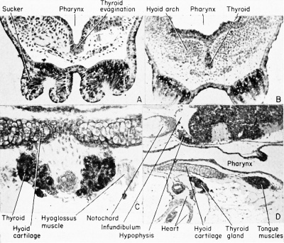
The thyroid is an endocrine gland which arises as a single median thickening and evagination from the floor of the pharynx between the base of the second pair of visceral arches, just before the time of
hatching and at about the 5 mm. stage. It later becomes a bi-lobed and solid organ, far removed from its site of origin. Only its original
lining is endodermal, the bulk of the gland being mesodermal in
origin. At about the 10 mm. stage, however, it becomes separated
from the pharyngeal floor as a closed sac. This divides into two lobular
and vesicular masses, and moves to a position on either side of
the hyoid cartilage apparatus. The thyroid gland of the 15 mm.
stage is divided but retains a connection at its anterior ends by a
short isthmus, so that it takes the shape of an inverted "Y," with a
short base. The wings move posteriorly, along the ventral face of
the hyoid cartilage, and these changes are correlated with gradual
changes in the hyoid apparatus. The glands enlarge, the surrounding cartilaginous elements expand, the major blood vessels become
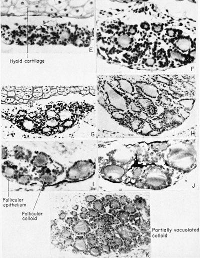
enmeshed in them, and eventually they come to lie close to the heart. At the forelimb emergence stage of metamorphosis there is a marked distension of the follicles, and by the tail resorption stage there is high epithelium liquefaction while erosion of the colloid and follicular collapse occur. At this time the genio-hyoid, hypoglossus, andsternohyoid muscles are in close proximity to the thyroid, a situation which does not persist to the adult. The gland shows hyperactivity during metamorphosis with relative inactivity at the completion of the metamorphic process. Its activity during these phases of development is closely correlated with the development and function of the pituitary gland, particularly of the basophilic cells in the pituitary gland. In later stages it may be seen attached to the ventral aspect of the hypoglossus muscle. The fully formed thyroid gland consists of separate follicles, each made up of a single circular layer of cuboidal (endodermal) epithelial cells, in the center of which is a lumen filled with a colloidal mass.
The Tongue
The tongue appears just before metamorphosis and is indicated as an elevation in the anterior floor of the pharynx, just posterior to the site of origin of the thyroid gland. Anterior to this the pharyngeal floor is depressed and glandular, but during metamorphosis this glandular area becomes the free anterior tip of the tongue.
The Lungs
Shortly after the time of hatching, when the larva measures about 6 mm. in length, there appear bifurcating but solid cell proliferations from the pharyngeal endoderm just behind the developing heart. These soon develop into paired saccular evaginations, directed posteriorly. These lung buds arise from the median ventral floor of the pharynx at about the level of the rudimentary sixth visceral (fifth branchial) pouch. The single short tubular connection of the lung buds opens into the foregut through the glottis. The connecting column of ceHs which has acquired a lumen, from which the lung buds arise, will become the trachea. At the level of the glottis there is a short transverse chamber known as the laryngeal chamber. The more posterior bi-lobed mass eventually will open up as the primary bronchi or lung buds. In the 1 1 mm. stage each lung bud will be surrounded by peritoneal epithelium and will be invested with splanchnic mesenchyme. Later, each lung will become an ovoid, thin-walled, and slightly alveolated sac, lined with endodermal squamous epithelium. Outside of this epithelium, and constituting the substance of the lung, are connective tissue, blood, and lymph vessels, all of mesodermal origin.
The Liver
The liver originates very early as a single median ventral endodermal diverticulum which is directed posteriorly between the heart rudiment and the yolk mass. The diverticulum enlarges slowly and its anterior wall will thicken, become folded, and finally branched, to form the liver proper. The ultimate lobes of the liver will retain their tubular connection with the original diverticulum as the hepatic duct. The original diverticulum will elongate as the bile duct leading to a terminal vesicle, the gallbladder, which becomes very large in the tadpole. All of these derivatives become invested with connective tissue and blood from the splanchnic mesoderm, but some of the adjoining yolk cells become the true hepatic cells.
The Pancreas
The pancreas arises as three rudiments at about the 9 mm. stage in a manner somewhat similar to the liver. The first rudiment appears as a single posteriorly directed ventral diverticulum of the bile duct at its point of entrance into the foregut. This diverticulum soon divides into two and the cellular elements grow around the bile duct to fuse into a single mass of spongy tissue anterior to the bile duct. Subsequently a third mass of similar tissue arises from the dorsal wall of the gut, and attains junction with the original masses, all three to form the much-lobulated pancreas. These three pancreatic rudiments then use the single pancreatic duct which retains its original connection with the gut, just posterior to the liver or hepatic duct. It marks the boundary between the foregut and the midgut. The cells of the islet of Langerhans arise early from endoderm.
The Oesophagus and Stomach
Digestive system and derivatives. (A) Before metamorphosis. (B) After metamorphosis, (Modified and redrawn from the Leuckart wall chart.)]]
After hatching, the undifferentiated portion of the foregut between the glottis and the bile duct elongates to become the oesophagus. During early larval development this part of the gut becomes temporarily occluded by an oesophageal plug of cells whose origin is unknown. Its function during its brief appearance may be to help direct any water and food from the mouth out over the gills. These early larvae require no external source of food as they are supplied with abundant yolk. The oesophageal plug disappears by the 9 to 10 mm. stage of development.
Beginning at about the 11 mm. stage, the oesophagus develops into
the very distensible organ of the adult while the stomach becomes
differentiated simply by a dilation of the next adjacent part of the
foregut. By the time of metamorphosis the stomach is somewhat more
distended than the oesophagus but it is otherwise indistinguishable from it. In the newly metamorphosed frog, however, it assumes a
transverse position, shifting from the earlier longitudinal position,
and has all the characteristic layers of any vertebrate gut. This includes an inner and glandular layer of mucosa, intermediate layers of
connective and muscular tissue, and an outer covering of serosa.
The Midgut
The midgut is that portion of the original archenteron which is found dorsal to the yolk mass as long as this mass exists, having a roof and sides one cell in thickness. The floor is the thick yolk endoderm. After the time of hatching the yolk is rapidly absorbed. Before the time of metamorphosis the heretofore undifferentiated tubular midgut becomes very much elongated into a doubly-coiled tube which may be nine to ten times as long as the body of the tadpole. During metamorphosis, and while the tadpole changes from an herbivorous (of the tadpole) to an omnivorous (of the frog) diet, this midgut (potential small intestine) shortens to about three times the length of the body, and its histology changes correspondingly. That portion of the midgut directly posterior to the stomach becomes the bent duodenum or duodenal loop. The small intestine, like the stomach, is lined with a glandular mucosa and is supplied with both circular and longitudinal (involuntary and smooth) muscles and an outer serosa. It is suspended within the body cavity by a thin but double layer of the peritoneal epithelium.
During the the earliest stages of development (2.5 mm. stage) there
may be seen a sub-notochordal or hypochordal rod of pigmented cells,
two or three cells in diameter, lying between the roof of the midgut
and the notochord. There is positional and structural evidence that
this column of cells is at some time associated with the roof of the
archenteron, from the level of the liver to the posteriorly placed
neurenteric canal. It becomes entirely free from gut and notochord by
the 4.5 mm. stage and disappears shortly after the time of hatching.
It has no known function. It may be of evolutionary and ontogenetic
significance only.
The Hindgut
This is the smallest portion of the original archenteron which lies posterior to the yolk mass, between it and the posterior body wall. The endoderm ventral to the closed blastopore evaginates to fuse with the invaginating proctodeal ectoderm to form the anal plate. This plate finally ruptures at about the 4 mm. stage to form the anus, only the inner portion of which is lined with endoderm. The rectum is the enlarged posterior end of the archenteron, and is therefore endodermally lined. This does not develop until the time of metamorphosis.
Dorsal to the rectum are a temporary extension of the archenteron,
developed in consequence of the presence of the neurenteric canal,
and the posterior growth of the notochord. This is the post-anal gut.
At the 5 mm. stage only a remnant of the neurenteric canal can be
identified as parallel rows of pigmented cells extending ventro-posteriorly from the hindgut. This is the region of the disappearing postanal gut. It represents the enteric portion of the temporary neurenteric
canal connecting the posterior ends of the neurocoel and archenteron.
It has no derivative in the adult, but has homologues in the development of most of the vertebrates.
That portion of the hindgut between the rectum and the anus
forms an ectodermally lined chamber known as the cloaca. Into this
chamber empty the paired urogenital ducts, to be developed later.
Before metamorphosis there appears a ventral evagination of the
cloaca which gives rise to the bladder. This ultimately bi-lobed and
elastic organ has no direct connection with the excretory ducts as do
the bladders of higher forms. However, it is considered to be a reservoir, by way of the cloaca, for excretory fluids. In the closely related
toads, which live in hot sand, it also may be a reservoir for water
storage and an aid in respiration.
Summary of Embryonic Development of the 11 mm Frog Tadpole
Endodermal Derivatives
The derivatives of the primary digestive tube, from anterior to posterior, are:
- Mouth — with the exception of that part from oral aperture to point of the internal nares (choanae) .
- Pharynx — dorso-ventrally flattened cavity into which mouth leads.
- Visceral clefts — junction of endodermal lining of visceral pouch with ectodermal invagination from side of head to form clefts associated with visceral pouches II, III, IV, and V. Pouch VI never forms a cleft. By the time the clefts are open they are covered by the operculum so that they lead into the opercular chamber ventro-laterally. External gills degenerating.
- Thyroid gland — now separated from floor of pharynx, dividing posteriorly into two lobes. Found just anterior to heart as paired pigmented masses.
- Thymus gland — derived from dorsal part of first and second visceral pouches (first degenerating) and migrating posteriorly to position near the ear.
- Parathyroid glands (epithelioid bodies) — from ventral portions of third and fourth visceral pouches. Difficult to locate.
- Ultimobranchial bodies — small pigmented masses at level of trachea ventral to sixth visceral pouches. Difficult to locate.
- Trachea and lungs — laryngo-tracheal groove from median ventral floor of pharynx near visceral clefts leads into tubular trachea which bifurcates into two primary bronchi. These expand posteriorly into lungs.
- Oesophagus — gut from laryngo-tracheal groove to stomach, bending behind liver.
- Stomach — identified by folded lining and thick muscular walls.
- Duodenum — from pyloric end of stomach to coiled intestine.
- Liver — highly vascularized and enlarged organ which incorporates vitelline veins. Original liver diverticulum becomes gallbladder with common bile duct (ductus choledochus) leading into dorsal wall of duodenum.
- Pancreas — within curvature of stomach, duct joining common bile duct.
- Intestine — two complete spiral coils posterior to duodenum. Cloaca as yet unformed but pronephric ducts open into posterior end of hindgut.
| Historic Disclaimer - information about historic embryology pages |
|---|
| Pages where the terms "Historic" (textbooks, papers, people, recommendations) appear on this site, and sections within pages where this disclaimer appears, indicate that the content and scientific understanding are specific to the time of publication. This means that while some scientific descriptions are still accurate, the terminology and interpretation of the developmental mechanisms reflect the understanding at the time of original publication and those of the preceding periods, these terms, interpretations and recommendations may not reflect our current scientific understanding. (More? Embryology History | Historic Embryology Papers) |
Reference
Rugh R. Book - The Frog Its Reproduction and Development. (1951) The Blakiston Company.
Cite this page: Hill, M.A. (2024, April 16) Embryology Book - The Frog Its Reproduction and Development 12. Retrieved from https://embryology.med.unsw.edu.au/embryology/index.php/Book_-_The_Frog_Its_Reproduction_and_Development_12
- © Dr Mark Hill 2024, UNSW Embryology ISBN: 978 0 7334 2609 4 - UNSW CRICOS Provider Code No. 00098G


